Understanding Myofascial Hip & Lateral Thigh Pain
Hip & Lateral Thigh Pain
Anatomy and Pain Pattern OverviewLateral hip and thigh pain is a common musculoskeletal complaint and a frequent reason for primary care and orthopedic consultation. The traditional diagnosis of "trochanteric bursitis" has been the default explanation for decades, yet imaging-based research has reshaped this picture: in a sonographic series of 877 patients with greater trochanteric pain syndrome, Long et al. (2013) found gluteal tendinopathy in roughly 50% of cases and isolated bursitis in only about 8% — most pain is generated by the gluteal tendons and surrounding soft tissues rather than by the bursa itself.
Myofascial trigger points in the gluteus medius, gluteus minimus, tensor fasciae latae, and piriformis are the most common musculoskeletal sources of lateral hip pain. The gluteus minimus deserves special attention because its referral pattern — extending from the lateral hip down the entire lateral thigh to the ankle — is the most common cause of pseudo-sciatica, leading to frequent misdiagnosis as lumbar radiculopathy.
The clinical distinction between myofascial hip pain and structural hip pathology is essential. True hip joint disease (osteoarthritis, labral tear, AVN) typically produces deep groin pain worsened by weight-bearing, with restricted internal rotation on examination. Myofascial lateral hip pain is located over or posterior to the greater trochanter, is exquisitely tender to palpation, and does not produce mechanical catching or locking. The two conditions can coexist, but treating the myofascial component often provides substantial relief even when structural changes are present.
In sonographic series of greater trochanteric pain syndrome, isolated bursitis is present in only a small minority of cases — gluteal tendinopathy and surrounding soft-tissue dysfunction account for the majority of findings.
Anatomy & Key Trigger Points
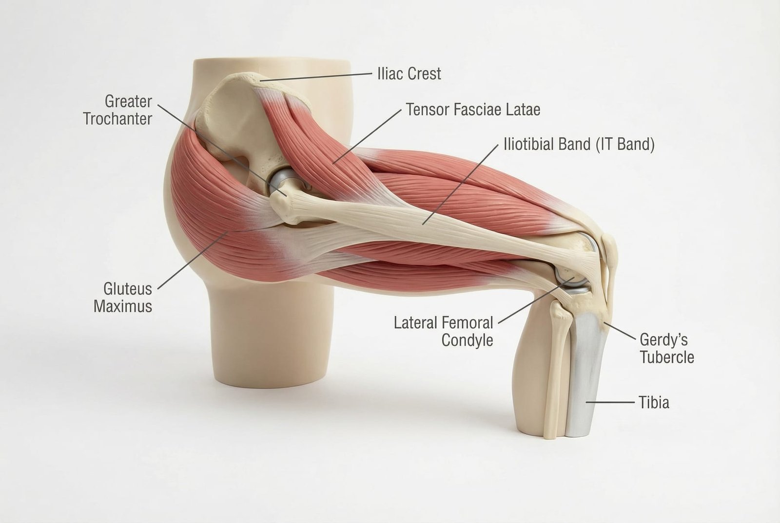
The lateral hip region contains multiple layers of muscle that work together to stabilize the pelvis during gait, control hip abduction, and maintain balance during single-leg stance. Trigger points in these muscles produce overlapping referral patterns that create the complex clinical presentation known as greater trochanteric pain syndrome.
Hip & Lateral Thigh Pain
The gluteus medius is the primary hip abductor and a critical stabilizer during single-leg stance and gait. Trigger points in this muscle are among the most common causes of lateral hip pain, producing referral along the posterior iliac crest, sacrum, and over the greater trochanter. Gluteus medius involvement contributes to many cases historically labeled "trochanteric bursitis" or "greater trochanteric pain syndrome," conditions in which gluteal tendinopathy and myofascial dysfunction are now recognized as far more common than true bursal inflammation (Long et al. 2013 found isolated bursitis in only ~8% of cases on ultrasound).
The gluteus minimus lies deep to the medius and is a hip abductor and internal rotator. Its trigger points produce one of the most clinically significant referral patterns in the body: pain extending from the lateral hip down the entire lateral thigh to the ankle, closely mimicking L5 radiculopathy. Gluteus minimus TrPs are the single most common cause of pseudo-sciatica in clinical practice, responsible for lateral thigh and leg pain that leads to unnecessary MRI and surgical consultations.
The TFL originates from the anterior superior iliac spine and inserts into the iliotibial band. Trigger points refer pain to the anterior and lateral hip, deep into the greater trochanter region, and occasionally down the lateral thigh along the ITB. TFL TrPs frequently coexist with gluteus medius involvement and contribute to the anterolateral component of "hip bursitis" presentations. Patients often report difficulty lying on the affected side at night.
The piriformis is a deep external rotator that lies in direct contact with the sciatic nerve. Trigger points produce deep buttock pain with referral to the posterior hip and down the posterior thigh. In hip pain presentations, piriformis TrPs contribute a posterior component that can be confused with posterior hip capsule pathology or SI joint dysfunction. Estimates of how often piriformis syndrome accounts for sciatica vary widely across the literature (commonly cited at roughly 0.3–6%), but in any individual case it should be considered alongside disc-related causes.
The largest and most powerful gluteal muscle. Trigger points in the gluteus maximus refer pain to the sacrum, coccyx, and deep buttock. Patients with gluteus maximus TrPs report an inability to sit comfortably, pain climbing stairs, and a deep ache in the buttock that worsens with prolonged hip flexion. These TrPs can mimic sacroiliac joint dysfunction and coccydynia and frequently coexist with gluteus medius and piriformis involvement.
The QL connects the last rib to the iliac crest and contributes to hip hiking and lateral trunk flexion. In the context of hip and lateral thigh pain, QL trigger points refer pain to the iliac crest, lateral hip, and greater trochanter region, overlapping significantly with gluteus medius referral. QL involvement should be suspected when hip pain extends superiorly to the flank or when patients report pain with side-bending.
The vastus lateralis is the largest of the quadriceps group, located on the lateral thigh. Trigger points refer pain along the lateral thigh from the greater trochanter to the lateral knee. Vastus lateralis TrPs can mimic or compound gluteus minimus referral and ITB syndrome, contributing to a continuous band of lateral thigh pain. Patients often describe difficulty walking downstairs and a deep ache that worsens after prolonged standing.
The iliopsoas complex (psoas major and iliacus) is the primary hip flexor. Trigger points refer pain to the anterior hip, groin, and deep anterior thigh. In hip pain presentations, iliopsoas TrPs contribute an anterior component that can mimic hip joint pathology, inguinal hernia, or femoral nerve entrapment. Patients with iliopsoas TrPs adopt an antalgic posture with the trunk flexed forward and the affected hip slightly flexed.
Gluteus Medius
Primary hip abductor referring pain across the lateral hip and iliac crest — the most common cause of pain misdiagnosed as trochanteric bursitis.
Gluteus Minimus
Deep hip abductor producing the classic pseudo-sciatica pattern: pain from the lateral hip to the ankle, mimicking L5 radiculopathy.
Tensor Fasciae Latae
Anterior hip muscle inserting into the ITB, referring pain to the anterolateral hip and along the lateral thigh.
Piriformis
Deep external rotator producing posterior hip and buttock pain with sciatic radiation — a contributor in a subset of sciatica presentations, with estimates varying widely across studies.
Referral Patterns
Understanding the distinct referral patterns of each gluteal and hip muscle is essential for accurate diagnosis. The gluteus minimus referral pattern is particularly important to recognize because it mimics lumbar radiculopathy so closely that it generates unnecessary MRI scans and surgical consultations.
Gluteus Medius
Gluteus Minimus
Tensor Fasciae Latae
Piriformis
Vastus Lateralis
Self-Assessment
The following clinical tests and self-assessment maneuvers can help identify myofascial trigger point contributors to hip and lateral thigh pain and differentiate them from structural hip pathology, true radiculopathy, and trochanteric bursitis.
Side-Lying Gluteus Medius Palpation
Deep Gluteus Minimus Palpation
Single-Leg Stand Test (Trendelenburg)
FABER/Patrick Test
Ober Test for ITB/TFL Tightness
Treatment Pathways
Effective management of gluteal myofascial pain combines targeted trigger point release, stretching, and progressive strengthening of the hip abductors. The rehabilitation component is critical — without restoring normal gluteus medius strength, recurrence is common because the muscle remains vulnerable to overload and reactivation of trigger points.
Treatment Pathways
Mechanism DiagramSelf-Care
- Foam rolling of the gluteus medius, TFL, and ITB — slow, sustained pressure for 60-90 seconds per region, avoiding direct pressure on the greater trochanter
- Tennis ball or lacrosse ball self-release for the gluteus minimus — lie on the ball against the wall, positioning it just above and anterior to the greater trochanter
- Piriformis stretch (figure-4 stretch) and gluteal stretching — hold 30 seconds, repeat 3 times, perform twice daily
- Sleep position modification — place a firm pillow between the knees to prevent hip adduction that compresses the lateral hip structures
- Avoid crossing legs, prolonged standing on one leg, and repetitive hip hiking — common perpetuating factors for gluteal TrPs
Manual Therapy
- Ischemic compression and sustained pressure release to gluteus medius, minimus, and TFL trigger points
- Deep tissue massage with specific focus on the posterior and lateral gluteal region, avoiding aggressive pressure directly over the trochanter
- Muscle energy techniques for the gluteus medius, piriformis, and TFL to restore normal muscle length and reduce TrP activity
- Myofascial release of the ITB and lateral fascial line to address fascial continuity patterns from hip to knee
- Joint mobilization of the hip and SI joint when hypomobility contributes to gluteal muscle overload
Interventional
- Dry needling of the gluteus medius and minimus TrPs — the most responsive gluteal muscles to needling with rapid symptom relief
- Trigger point injection with lidocaine into the gluteus medius and TFL for immediate pain reduction
- Ultrasound-guided dry needling of the gluteus minimus — deep location benefits from image guidance to ensure accurate needle placement
- Corticosteroid injection to the trochanteric bursa only when true bursitis is confirmed on imaging — most "bursitis" is actually myofascial
Rehabilitation
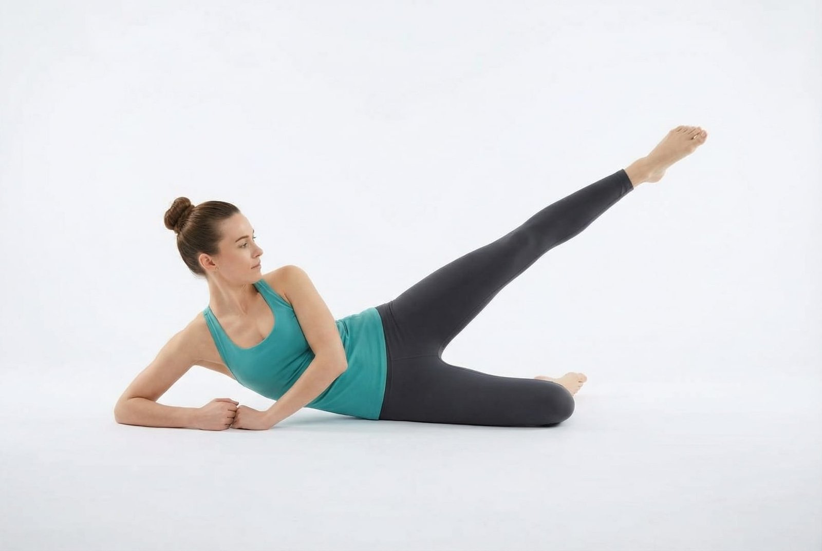
- Progressive hip abductor strengthening — side-lying hip abduction, clamshells, and standing hip abduction with resistance bands
- Single-leg balance training to restore gluteus medius neuromuscular control and pelvic stability during gait
- Gait retraining with emphasis on eliminating Trendelenburg pattern and reducing excessive hip adduction during stance phase
- Core stability training with emphasis on the lateral stabilization chain — gluteus medius, quadratus lumborum, and lateral trunk muscles
- Gradual return to provocative activities (side-lying sleep, prolonged walking, stair climbing) as symptoms resolve
Red Flags — When to See a Doctor
Before attributing hip and lateral thigh pain to a myofascial source, always screen for the following red flags that indicate potentially serious underlying pathology requiring urgent evaluation.
The gluteus medius is one of the most common myofascial contributors to lateral hip pain. Imaging studies (e.g., Long et al. 2013) show that isolated trochanteric bursitis accounts for only a small fraction of cases historically labeled "bursitis" — most are driven by gluteal tendinopathy with or without coexisting myofascial dysfunction.
Gluteus minimus trigger points produce the classic pseudo-sciatica pattern: pain extending from the lateral hip down the entire lateral thigh to the ankle. This pattern leads to unnecessary MRI and surgical consultations when the true source is muscular.
The key differentiator between true radiculopathy and gluteal pseudo-sciatica is the presence of neurological findings. Myofascial referral does not produce dermatomal sensory loss, reflex changes, or progressive motor weakness — findings that mandate spinal imaging.
Sleep disruption is a hallmark of gluteal myofascial pain — patients cannot lie on the affected side due to compression of TrPs against the mattress. Pillow placement between the knees and mattress assessment are essential parts of the treatment plan.
Treatment should combine self-release (foam rolling, tennis ball), targeted stretching, and progressive hip abductor strengthening. Dry needling of the gluteus medius and minimus is highly effective for resistant cases.
Always consider the gluteal muscles before ordering advanced imaging for lateral hip or thigh pain — a thorough palpation exam takes 5 minutes and can prevent months of unnecessary investigation.