Understanding Myofascial Heel Pain
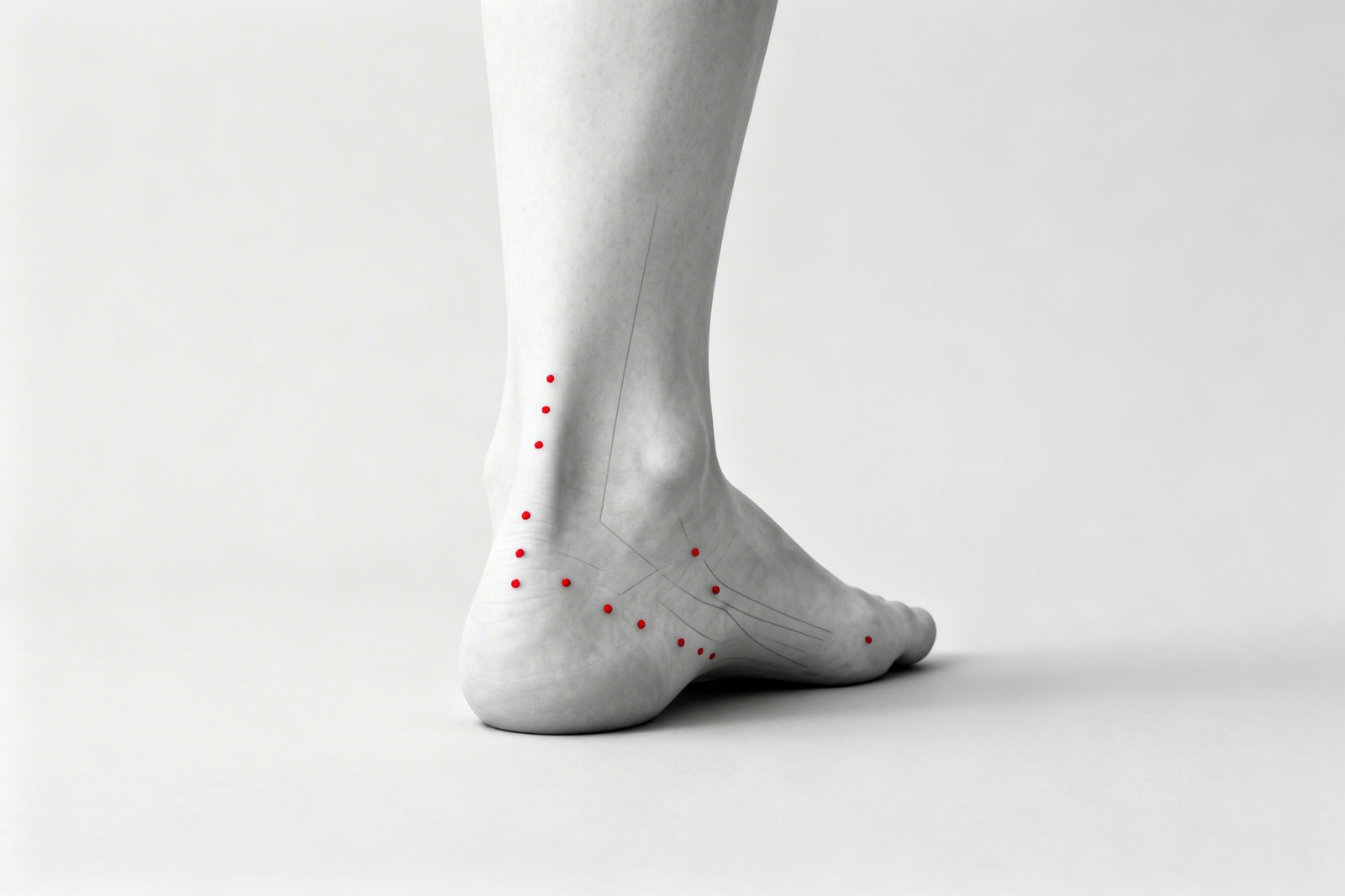
Heel Pain & Plantar Fasciitis Mimics
Anatomy and Pain Pattern OverviewPlantar fasciitis is the most commonly diagnosed cause of heel pain, affecting roughly a tenth of the general population over a lifetime. Standard treatment — stretching, orthotics, night splints, and corticosteroid injections — leads to resolution in the majority of patients within 12 months, with a substantial proportion improving regardless of specific management strategy. But what about the meaningful subset who fail to improve? An emerging body of evidence points to myofascial trigger points in the calf muscles and intrinsic foot muscles as important unrecognized pain generators in treatment-resistant heel pain.
The soleus, gastrocnemius, quadratus plantae, and flexor digitorum brevis all refer pain to the heel and sole of the foot in patterns that are clinically indistinguishable from plantar fasciitis based on pain location alone. Clinical examination consistently identifies myofascial trigger points in the soleus and surrounding muscles as common contributors in patients with recalcitrant plantar heel pain — trigger points that often resolve with targeted treatment, finally ending months or years of unsuccessful fascial therapy.
A meaningful proportion of patients with treatment-resistant plantar fasciitis have soleus trigger points contributing to their pain — a treatable condition that often responds well to targeted intervention.
The distinction between fascial and myofascial heel pain is more than academic — it determines the treatment approach. True plantar fasciitis responds to fascial stretching, orthotics, and load modification. Myofascial heel pain responds to trigger point release in the calf and intrinsic foot muscles, eccentric calf strengthening, and addressing perpetuating factors like footwear and gait mechanics. When both sources are present, both must be addressed for complete resolution.
Anatomy & Key Trigger Points
The heel and sole of the foot receive referred pain from multiple muscles in the posterior leg compartment and the intrinsic foot muscles. Understanding which muscles refer to which specific location is essential for identifying the true pain generator when standard plantar fasciitis treatment fails.
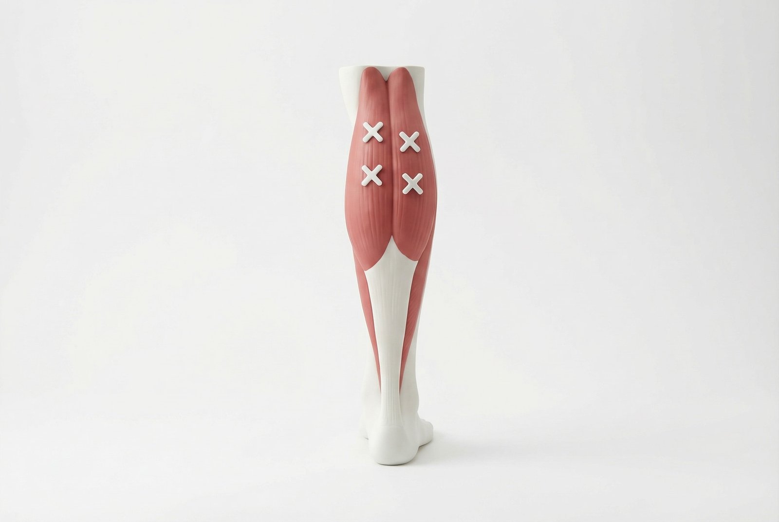
Heel Pain & Plantar Fasciitis Mimics
Gastrocnemius
Soleus
Quadratus Plantae (Flexor Accessorius)
Abductor Hallucis
Tibialis Posterior
Flexor Digitorum Longus
Flexor Hallucis Longus
Flexor Digitorum Brevis
Soleus
The most common myofascial mimic of plantar fasciitis — refers pain to the posterior and plantar heel, often missed because the source is in the calf.
Gastrocnemius
Superficial calf muscle referring pain to the medial arch and instep — extends the pain zone beyond the classic heel-only pattern of fasciitis.
Quadratus Plantae
Deep intrinsic foot muscle referring pain to the exact calcaneal insertion site — clinically indistinguishable from plantar fasciitis by location alone.
Tibialis Posterior
Deep posterior leg muscle referring pain to the Achilles region, heel, and arch — creates a broad pattern overlapping both fasciitis and tendinopathy.
Referral Patterns
The referral patterns of the calf and intrinsic foot muscles create a complex overlap with plantar fasciitis pain. The key diagnostic principle is that myofascial heel pain is reproduced by palpating trigger points in the muscle bellies (in the calf or sole of the foot), not at the fascial insertion on the calcaneus.
Soleus
Gastrocnemius (Medial Head)
Quadratus Plantae
Tibialis Posterior
Abductor Hallucis
Self-Assessment
The following tests can help identify myofascial trigger point contributors to heel pain and differentiate them from true plantar fasciitis. The hallmark of myofascial heel pain is reproduction of the heel symptoms by palpating trigger points in the calf muscles — demonstrating that the pain source is proximal, not at the foot.
Soleus Deep Palpation Test
Gastrocnemius Squeeze Test
Plantar Intrinsic Muscle Palpation
Windlass Test (Fascial vs. Muscular)
First-Step Pain Characterization
Treatment Pathways
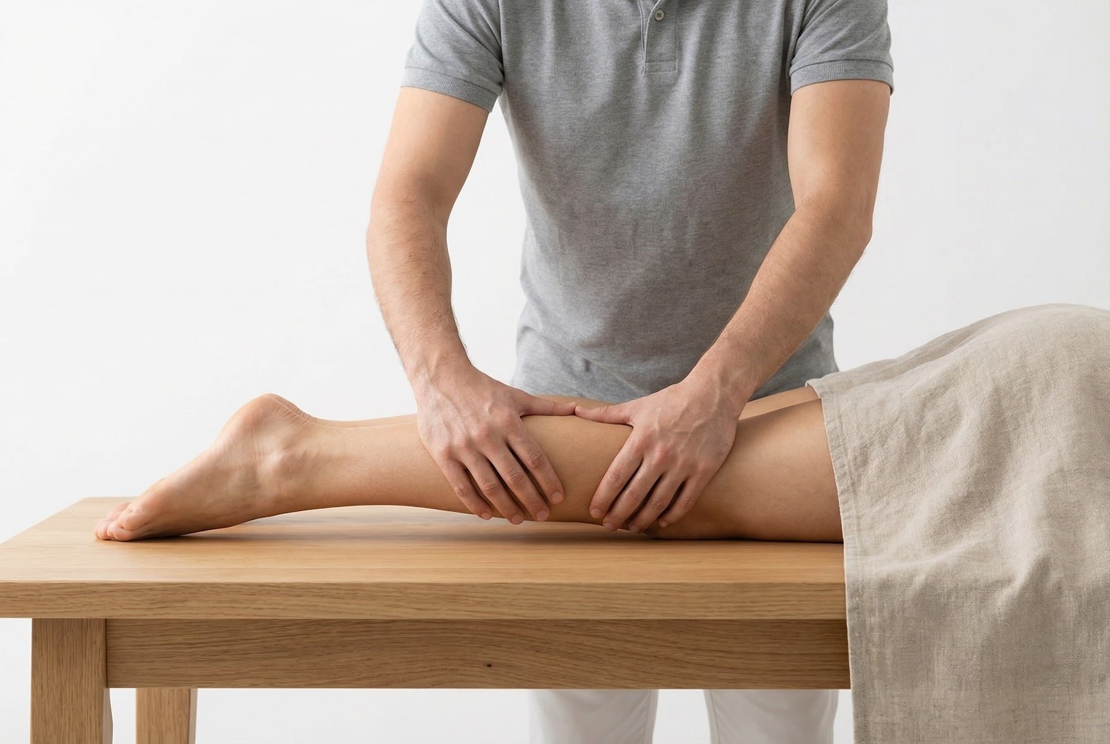
Treatment of myofascial heel pain focuses on releasing trigger points in the calf muscles and intrinsic foot muscles, followed by eccentric calf strengthening and gradual return to weight-bearing activities. The key insight is that treating calf tightness often improves the heel pain — a concept counterintuitive to patients accustomed to treating the foot itself.
Treatment Pathways
Mechanism DiagramSelf-Care
- Calf stretching — sustained wall stretch with the knee straight (gastrocnemius) and bent (soleus), hold 30 seconds, 3 repetitions each, 3 times daily
- Tennis ball or frozen water bottle rolling under the sole of the foot — 5 minutes of slow, sustained pressure targeting the muscle bellies, not just the fascia
- Self-release of the soleus using a foam roller or lacrosse ball — sit with the calf on the roller, cross the opposite leg on top for added pressure, roll slowly
- Night splint or Strassburg sock to maintain ankle dorsiflexion during sleep — stretches both the calf muscles and plantar structures
- Evaluate footwear — transition from flat shoes or flip-flops to shoes with moderate heel elevation and arch support to reduce calf and plantar muscle loading
Manual Therapy
- Deep tissue release of the soleus and gastrocnemius trigger points — ischemic compression with sustained pressure for 60-90 seconds per TrP
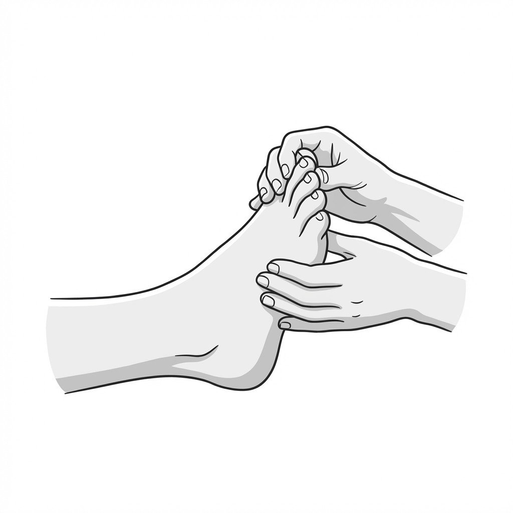
- Plantar intrinsic muscle release — sustained pressure to the quadratus plantae, abductor hallucis, and flexor digitorum brevis through the sole of the foot
- Myofascial release of the posterior fascial chain from the calf through the plantar fascia to the toe flexors
- Ankle and subtalar joint mobilization when restricted dorsiflexion contributes to calf overload
- Instrument-assisted soft tissue mobilization (IASTM) along the Achilles tendon, plantar fascia, and intrinsic foot muscles
Interventional
- Dry needling of the soleus trigger points — supported by emerging evidence as an effective interventional option for myofascial heel pain, with many patients reporting meaningful symptom relief
- Dry needling of the gastrocnemius and tibialis posterior TrPs for comprehensive posterior chain treatment
- Trigger point injection with local anesthetic into accessible calf and intrinsic foot muscle TrPs
- Extracorporeal shockwave therapy (ESWT) — evidence supports efficacy for both plantar fasciitis and myofascial trigger points in the calf and plantar muscles
Rehabilitation
- Eccentric calf strengthening — Alfredson protocol (heel drops off a step) for both straight-knee (gastrocnemius) and bent-knee (soleus) positions
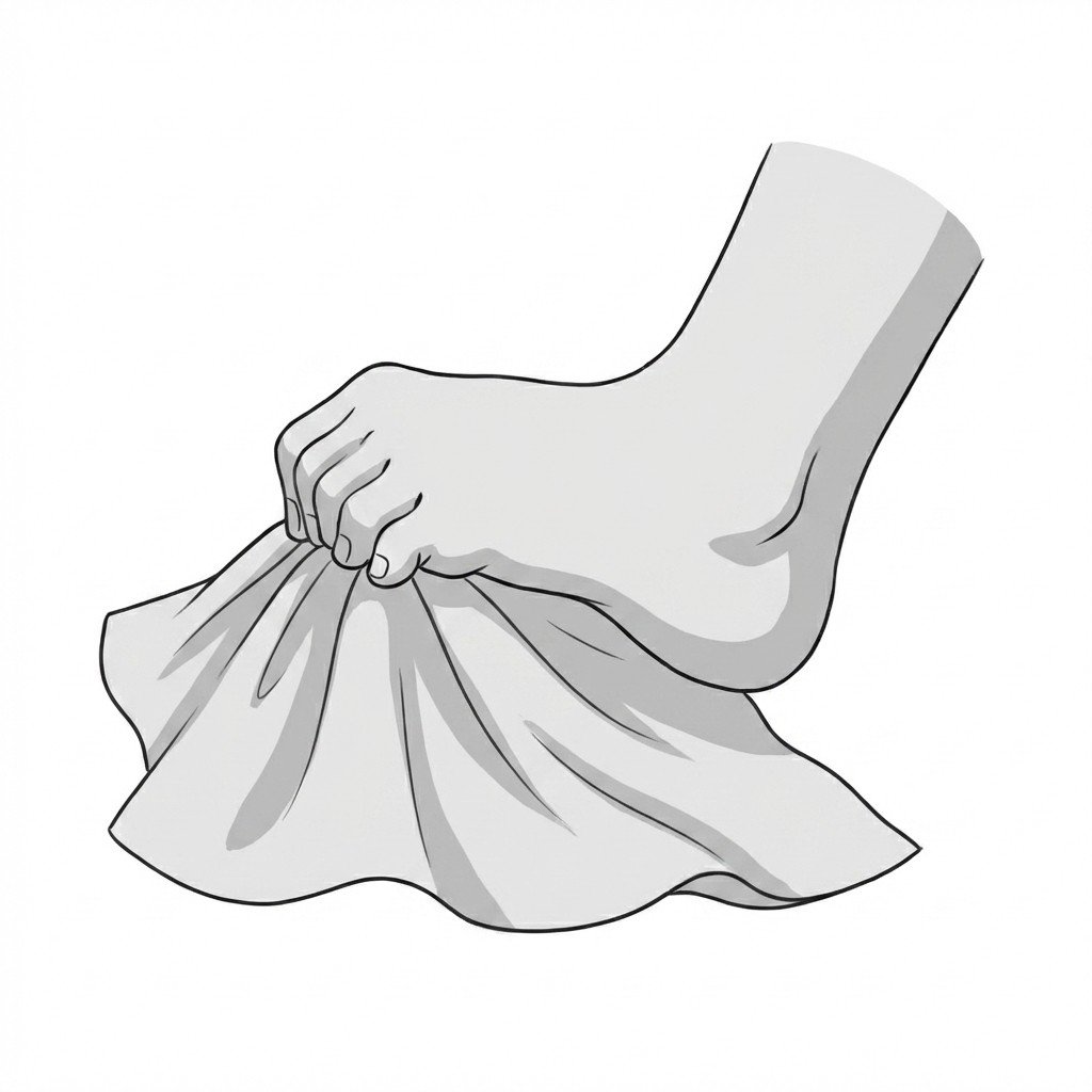
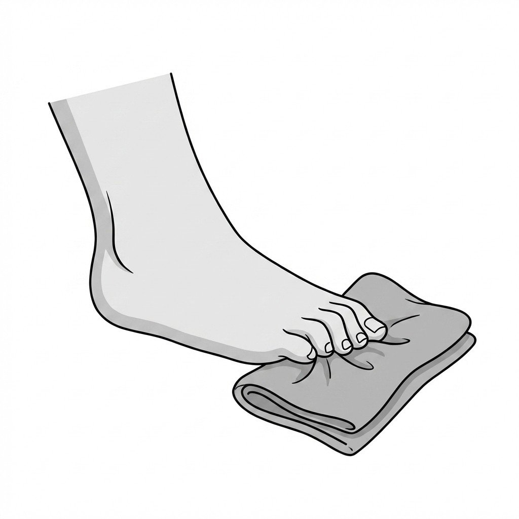
- Intrinsic foot muscle strengthening — towel curls, marble pickups, and short foot exercise to restore dynamic arch support
- Progressive weight-bearing activity — graduated walking program with appropriate footwear, increasing duration by 10% per week
- Balance and proprioceptive training on unstable surfaces to improve ankle stability and reduce compensatory muscle overload
- Gait analysis and correction of overpronation, overstriding, or forefoot strike patterns that perpetuate calf and plantar muscle overload
Red Flags — When to See a Doctor
Before attributing heel pain to a myofascial source, always screen for the following red flags indicating potentially serious underlying pathology requiring urgent evaluation.
The soleus is widely considered the most common myofascial mimic of plantar fasciitis, referring pain directly to the plantar and posterior heel. A meaningful proportion of patients diagnosed with plantar fasciitis who fail standard treatment have unrecognized soleus trigger points contributing to their pain.
The quadratus plantae — a small intrinsic foot muscle — refers pain to the exact calcaneal insertion site attributed to plantar fasciitis. This muscle is almost never examined in standard clinical evaluations, making it a critical overlooked source of heel pain.
The Windlass test can help differentiate fascial from muscular heel pain. A negative Windlass test (no pain with great toe dorsiflexion) in a patient with heel pain strongly suggests a myofascial rather than fascial source.
When heel pain does not improve with standard plantar fasciitis treatment (stretching, orthotics, night splints), the calf muscles should be the next evaluation target. Palpating the soleus and gastrocnemius for trigger points that reproduce the heel pain is the key diagnostic step.
Treatment should address the entire posterior chain — calf muscles, Achilles complex, and plantar intrinsic muscles — rather than focusing solely on the plantar fascia. Eccentric calf strengthening combined with trigger point release produces the best long-term outcomes.
Always screen for red flags before attributing heel pain to a myofascial source — calcaneal stress fracture, peripheral neuropathy, tarsal tunnel syndrome, DVT, and Achilles rupture all require specific evaluation and management.
Movement therapy
Exercises for Plantar Fasciitis
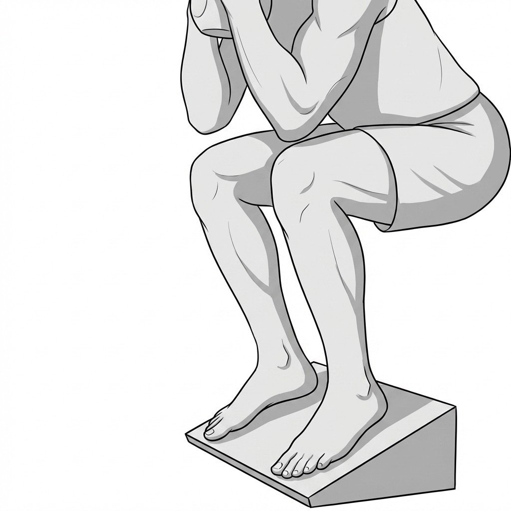
These exercises address the calf tightness and intrinsic foot weakness that perpetuate plantar heel pain — calf stretching on a slant reduces morning tensile load on the fascia, while toe yoga and towel scrunches restore the small-muscle control that offloads the plantar structures. Start with 2–3 and progress as tolerated.