§ 01

What Are Fascial Plane Blocks?

Fascial Plane Blocks

Fascial Plane Blocks

Overview Diagram

Fascial plane blocks are a category of ultrasound-guided regional anesthesia techniques in which local anesthetic is deposited between layers of fascia to influence the sensory nerves that travel through these tissue planes. Originally developed mainly for post-surgical analgesia, these blocks are increasingly being explored in chronic pain conditions, including selected cases of thoracic, chest wall, and lumbar myofascial pain.

The idea is anatomically elegant: muscles are enclosed within fascial sheaths, and the nerves supplying these muscles and their overlying tissues travel through predictable compartments. By injecting local anesthetic into these compartments under ultrasound guidance, a single injection may influence multiple segmental nerves simultaneously, providing broader regional analgesia than one local trigger point injection.

Fascial plane blocks use the anatomy of tissue compartments to create regional analgesia from a single ultrasound-guided injection.

For patients with myofascial pain syndrome, fascial plane blocks serve a specific role: they may provide temporary regional analgesia that reduces the pain–spasm cycle and creates a window in which physical therapy, stretching, and functional rehabilitation become easier to perform.

§ 02

Types of Fascial Plane Blocks

Fascial Plane Block Types

Fascial Plane Block Types

Comparison Chart

Erector Spinae Plane (ESP) Block

Target: Deep to erector spinae muscle, superficial to transverse processCoverage: Variable thoracic or lumbar segmental spread depending on level, volume, and anatomyVolume: Often 20–30 mL of dilute ropivacaine or bupivacaine, adjusted to context and safety limits
  • Local anesthetic may spread cranio-caudally along the erector spinae fascial plane, potentially covering multiple segmental levels with a single injection
  • Often used to influence posterior thoracic or lumbar pain through spread toward dorsal rami and related posterior structures, though exact spread is variable
  • Technically straightforward in experienced hands because the transverse process is usually an identifiable ultrasound landmark
  • Generally viewed as having a favorable safety profile compared with deeper paravertebral or neuraxial techniques, while still requiring procedural caution
  • Widely adopted since 2016 for truncal analgesia, with chronic pain applications now expanding but still less established than post-surgical use
Best for: Selected chronic thoracic and lumbar myofascial pain patterns, post-thoracotomy pain, and some posterior truncal pain syndromes

Pectoral Nerve Blocks (Pecs I & Pecs II)

Target: Pecs I: between pectoralis major and minor. Pecs II: between pectoralis minor and serratus anteriorCoverage: Lateral and medial pectoral nerves, intercostobrachial nerve, and variable anterior-lateral chest wall coverageVolume: Pecs I: often around 10 mL; Pecs II: often around 20 mL of dilute local anesthetic, adjusted to patient and procedure
  • Pecs I targets the fascial plane between pectoralis major and minor, aiming to influence the lateral and medial pectoral nerves
  • Pecs II extends coverage toward the axilla and lateral chest wall by depositing local anesthetic deep to pectoralis minor
  • Can be useful for chest wall pain patterns that do not respond adequately to manual therapy, exercise, and more local treatments
  • May be considered in patients with anterior chest wall pain patterns that have already been medically evaluated for more serious causes
  • Usually performed supine with the arm positioned for access, with relatively straightforward ultrasound visualization of pectoral fascial layers
Best for: Selected anterior chest wall myofascial pain, pectoralis trigger points, and post-breast-surgery pain patterns

Serratus Anterior Plane (SAP) Block

Target: Superficial or deep to serratus anterior muscle at the mid-axillary lineCoverage: Lateral cutaneous branches of intercostal nerves with variable lateral thoracic wall spreadVolume: Often 20–40 mL of dilute ropivacaine or bupivacaine depending on indication and dosing limits
  • Aims to provide analgesia to the lateral thoracic wall by blocking the lateral cutaneous branches of the intercostal nerves
  • Two approaches are commonly described: superficial (above serratus anterior) and deep (between serratus anterior and external intercostal tissues)
  • The superficial approach is often considered easier and may provide broader cutaneous spread, though both approaches are used
  • Can be a reasonable option for suspected serratus anterior or lateral chest wall myofascial pain that is difficult to manage with standard approaches
  • Generally lower pneumothorax risk than direct intercostal nerve blocks when performed carefully with ultrasound and tangential needle control
Best for: Selected lateral chest wall myofascial pain, serratus-related pain, and rib-fracture-associated pain

Quadratus Lumborum (QL) Block

Target: Adjacent to the quadratus lumborum muscle (anterior, lateral, or posterior approach)Coverage: Variable thoracolumbar abdominal wall and flank coverage depending on approach and spreadVolume: Often 20–30 mL of dilute ropivacaine or bupivacaine, adjusted to indication and total dose limits
  • Three commonly described approaches — QL1 (lateral), QL2 (posterior), and QL3 (anterior/transmuscular) — each with somewhat different spread patterns
  • The anterior (QL3) approach is often discussed as producing broader and more consistent spread in some studies, though results vary
  • Local anesthetic may spread along the thoracolumbar fascia and in some cases toward the paravertebral region, contributing to broader somatic analgesia
  • Can be particularly useful for deep quadratus lumborum-related pain patterns that are difficult to access with standard trigger point techniques
  • May provide several hours of analgesia, creating a temporary window to reduce guarding and improve participation in rehabilitation
Best for: Selected flank and lower back myofascial pain, quadratus lumborum-related pain, and abdominal wall pain
§ 03

Mechanism of Action

Fascial plane blocks likely work through a combination of peripheral nerve blockade, regional sensory modulation, and temporary reduction of ongoing nociceptive input. The key to their usefulness is the anatomy of fascial planes, which can allow local anesthetic to spread to multiple neural targets from one injection point.
Fascial Plane Blocks

Fascial Plane Blocks

Mechanism Diagram

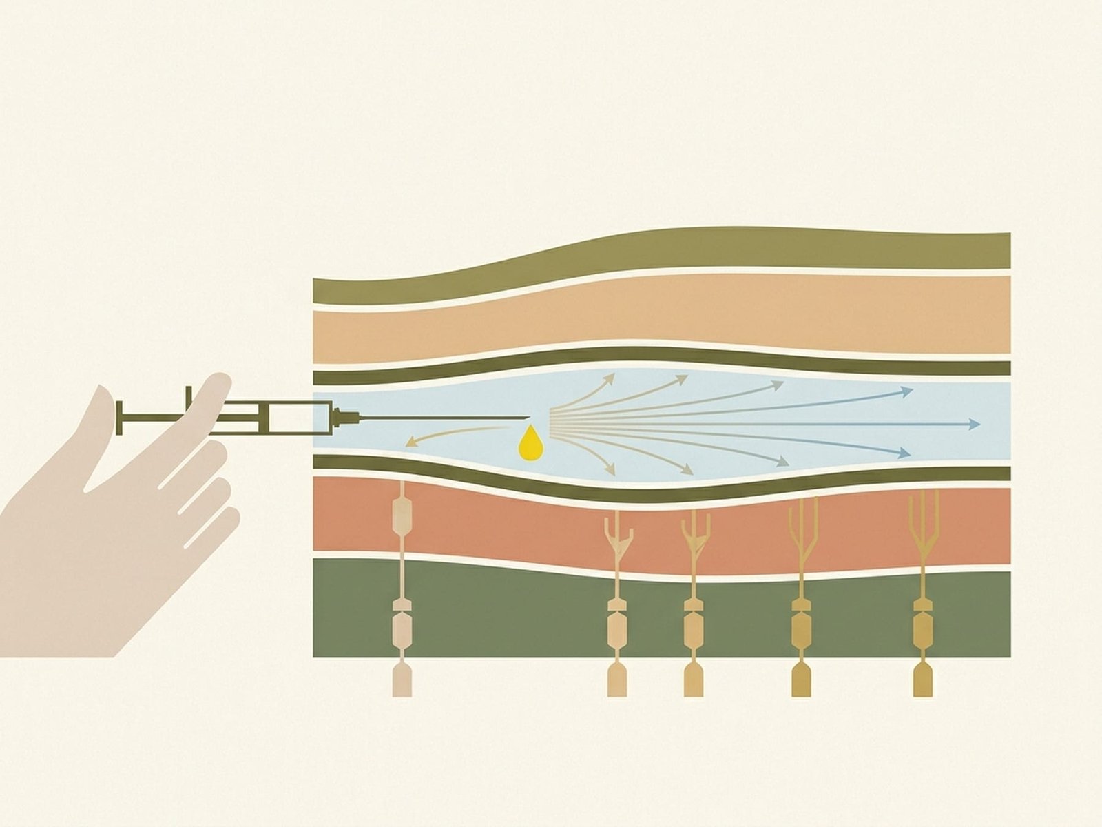
Fascial Plane Spread of Local Anesthetic

Local anesthetic deposited between fascial layers can spread along natural tissue planes. This allows a single injection to potentially influence multiple sensory nerves traversing the compartment, providing broader regional analgesia than a single point-specific injection.

Sympathetic Modulation

Some fascial plane blocks — particularly ESP and certain QL approaches — may produce partial sympathetic effects when local anesthetic spreads toward deeper paravertebral structures. This is best described as a possible contributing mechanism rather than a guaranteed effect in every case.

Sensory Nerve Blockade

Local anesthetic molecules bind to voltage-gated sodium channels on sensory nerve fibers traversing the fascial plane, reducing depolarization and action-potential propagation. This decreases nociceptive transmission from the affected region and can provide meaningful regional analgesia.

Reduction of Ongoing Nociceptive Input

Chronic myofascial trigger points can maintain a persistent stream of nociceptive input into the nervous system. Fascial plane blocks may temporarily interrupt that input, which can reduce pain amplification and create a window in which movement, stretching, and therapy become easier to tolerate.
How Fascial Plane Blocks Work
Fascial Plane Spread

Local anesthetic may spread along natural tissue planes to reach multiple nerve branches from one injection point.

Sympathetic Modulation

Some blocks may produce partial sympathetic effects when spread reaches deeper adjacent structures.

Sensory Nerve Blockade

Sodium-channel blockade of sensory nerve fibers traversing the fascial compartment reduces nociceptive transmission.

Nociceptive Reset Window

Temporary interruption of ongoing nociceptive input can make movement and rehabilitation more tolerable.

§ 04

Ultrasound-Guided Technique

Ultrasound Scanning & Landmark Identification

A high-frequency linear ultrasound probe (or curvilinear probe for deeper blocks) is used to identify the relevant anatomical landmarks. For ESP blocks, the transverse process is identified; for Pecs blocks, the pectoral fascial layers; for SAP blocks, the serratus anterior and ribs at the mid-axillary line; for QL blocks, the quadratus lumborum muscle and its fascial boundaries.

Patient Positioning & Sterile Preparation

The patient is positioned to optimize ultrasound visualization and needle access. ESP and QL blocks are typically performed in the prone or lateral decubitus position. Pecs and SAP blocks are performed supine with arm abduction. The skin is prepared with chlorhexidine antiseptic and draped. A sterile ultrasound probe cover is applied.

Needle Insertion Under Real-Time Ultrasound Guidance

A 22-gauge, 80–100 mm echogenic block needle is advanced in-plane with the ultrasound beam for continuous tip visualization. The needle is directed toward the target fascial plane while monitoring for correct trajectory and avoiding adjacent structures such as pleura, vessels, and deeper neural structures.

Hydrodissection & Confirmation of Fascial Plane

A small test bolus of normal saline or dextrose 5% (1–2 mL) is injected to confirm correct needle tip position within the fascial plane. Correct placement is suggested by visualizing separation of fascial layers on ultrasound. If the injectate does not spread as expected, the needle is repositioned.

Local Anesthetic Injection

Once correct fascial plane placement is confirmed, the planned volume of local anesthetic is injected incrementally with intermittent aspiration to reduce the risk of intravascular injection. Real-time ultrasound visualization helps confirm spread along the intended fascial plane.

Post-Procedure Assessment

The patient is monitored after the procedure. Sensory testing may be used to estimate block effect, and the patient is observed for complications such as local anesthetic systemic toxicity (LAST), pleural irritation or pneumothorax for thoracic blocks, or hemodynamic changes when broader spread is suspected. Pain scores and functional response are documented before and after the procedure.
§ 05

Clinical Evidence

Emerging Evidence for MPSSelected refractory thoracic and lumbar myofascial pain

Forero et al. (2016)

This landmark case series described the erector spinae plane block for thoracic neuropathic pain and helped stimulate wider adoption of the technique. It established clinical feasibility and opened the door to later chronic pain applications, including possible use in selected myofascial pain presentations.

Adhikary et al. (2018)

Review work on ESP block applications suggested effective analgesia across multiple indications including chronic thoracic pain, post-surgical pain, and rib fractures. Cadaveric and imaging studies supported spread toward deeper structures, though exact patterns remain variable.

Chin et al. (2017)

This work helped support QL block use for thoracolumbar and abdominal wall analgesia, with the transmuscular (anterior) approach often discussed as producing broader and more reliable spread in some settings.

Case Series — Refractory Myofascial Pain

Small case series and reports suggest that some patients with chronic thoracic and lumbar myofascial pain syndrome refractory to trigger point injections, dry needling, and physical therapy may achieve meaningful short-term pain reduction after fascial plane blocks. Duration of benefit appears variable, ranging from hours to days and sometimes longer.

Post-Surgical Pain Meta-Analyses

The strongest evidence base for fascial plane blocks remains post-surgical analgesia — particularly ESP blocks after thoracic or spinal surgery, and Pecs blocks after breast surgery. These studies support the broader analgesic plausibility of the techniques, but they are not the same as direct evidence for chronic MPS.

Emerging MPS-Specific Research

Prospective studies specifically evaluating fascial plane blocks for myofascial pain syndrome are still limited. Early reports are promising, especially when the blocks are integrated with rehabilitation rather than used alone, but the evidence remains emerging rather than definitive.

Meaningful Relief in Some Cases

Case reports and small series suggest clinically relevant short-term pain reduction in selected refractory patients

Block Duration

Single injection often provides hours of regional analgesia, sometimes longer depending on technique and drug

Generally Favorable Safety Profile

Serious complications appear uncommon when blocks are performed under ultrasound guidance by trained clinicians
§ 06

Ideal Candidates

Refractory Thoracic or Lumbar Myofascial Pain

Patients with chronic MPS of the thoracic or lumbar paraspinal muscles who have failed conservative treatments including physical therapy, manual therapy, dry needling, and trigger point injections. Fascial plane blocks offer a regional approach when point-specific treatments are insufficient.

Widespread Multi-Level Trigger Points

Patients with trigger points spanning multiple segmental levels. A regional block may be more practical than numerous individual trigger point injections when the pain pattern is broad and regionally clustered.

Chest Wall Myofascial Pain Syndromes

Patients with pectoralis, serratus anterior, or intercostal-region myofascial pain that is difficult to treat with standard approaches. Pecs blocks and SAP blocks may target these regions through their fascial compartments.

Pain–Spasm Cycle Breaking

Patients with severe muscle guarding and spasm overlying trigger points, making direct treatment difficult or poorly tolerated. A fascial plane block may provide a temporary analgesic window for physical therapy and stretching.

Concurrent Neuropathic and Myofascial Pain

Patients with mixed pain presentations — both myofascial trigger points and underlying neuropathic pain components — may sometimes benefit when a regional technique addresses more than one contributor at once.
§ 07

Contraindications & Side Effects

Absolute Contraindications

Relative Contraindications

Side Effects

Generally Favorable Safety Profile

Fascial plane blocks are generally considered safer than neuraxial techniques when performed by trained proceduralists under ultrasound guidance, but they still require careful consent, dosing, and monitoring. Serious complications are uncommon, not impossible.

Temporary Motor Weakness

Depending on the block type, spread pattern, and local anesthetic concentration, temporary weakness of nearby muscles may occur. This is usually transient and resolves as the block wears off.

Hypotension (Occasional)

ESP and QL blocks that produce broader spread may occasionally contribute to mild hypotension, particularly with bilateral techniques or in more sensitive patients. Monitoring is appropriate.

Injection Site Soreness

Mild tenderness at the needle insertion site is common for 1–3 days after the procedure. This is related to needle passage through soft tissues and usually resolves spontaneously.

Pneumothorax (Rare)

Thoracic fascial plane blocks carry a small pneumothorax risk if the needle advances beyond the intended plane toward the pleura. Real-time ultrasound guidance and continuous tip visualization are used to minimize this risk.

Local Anesthetic Systemic Toxicity (Rare but Serious)

LAST can occur with inadvertent intravascular injection or excessive systemic absorption. Symptoms may include metallic taste, tinnitus, perioral numbness, seizures, or cardiac instability. Appropriate rescue protocols and lipid emulsion availability are essential.
§ 08

Fascial Blocks vs Other Treatments

Comparison
Fascial Block AdvantageFascial Block Limitation

vs Trigger Point Injections

Broader regional coverage from a single injection; may influence multiple trigger points simultaneously
Less precise for one specific trigger point; requires ultrasound equipment and procedural expertise

vs Dry Needling

May provide hours of meaningful regional analgesia and reduce guarding without depending on a local twitch response
More resource-intensive, uses larger injectate volumes, and requires post-procedure monitoring

vs Epidural Steroid Injection

Avoids the neuraxis and therefore avoids some neuraxial-specific risks such as dural puncture or epidural hematoma
Does not create the same central neuraxial block and is usually limited to truncal or regional targets

vs Oral Analgesics

Targeted regional effect without the burden of chronic systemic medication exposure
Requires a procedural setting; benefit is usually temporary and should not be oversold as a stand-alone solution
§ 09

Expected Timeline

15–30 m

Block OnsetSensory change often develops within about 15 to 30 minutes after injection, though onset varies

Hours

Analgesic DurationA single injection commonly provides several hours of regional analgesia and sometimes longer depending on anesthetic choice and spread

Variable

Treatment SeriesSome chronic MPS patients may benefit from repeated blocks combined with physical therapy, but there is no single fixed series that fits every case
Key Takeaways
  1. Fascial plane blocks are ultrasound-guided regional anesthesia techniques that deposit local anesthetic between fascial layers, providing broader regional analgesia for selected truncal myofascial pain syndromes.

  2. Four commonly discussed block types for these pain regions are ESP block, Pecs I/II, SAP block, and QL block.

  3. The mechanism likely involves local anesthetic spread along fascial planes to sensory nerves, with possible additional sympathetic and central-modulation effects in some cases.

  4. Direct evidence for MPS is still emerging. The strongest evidence base remains post-surgical analgesia, while chronic myofascial pain evidence is mainly early-stage and case-series based.

  5. These blocks can create a temporary analgesic window that makes physical therapy, stretching, and rehabilitation easier to tolerate in selected patients.

  6. Fascial plane blocks require a trained interventionalist with ultrasound proficiency and should be integrated into a multimodal plan, not treated as a stand-alone cure.

  7. The safety profile is generally favorable compared with neuraxial techniques, but anticoagulation status, dosing limits, and post-procedure monitoring still matter.