Understanding Myofascial Pelvic Pain
Chronic Pelvic Pain
Anatomy and Pain Pattern OverviewChronic pelvic pain (CPP) is common and often frustrating to evaluate. Even after workups that may include imaging, cystoscopy, laparoscopy, or laboratory studies, many patients are left without a single clear structural explanation. This diagnostic gap is one reason myofascial trigger points are increasingly recognized as an important and frequently overlooked contributor to chronic pelvic pain.
Myofascial trigger points in the pelvic floor muscles, obturator internus, piriformis, lower abdominals, and iliopsoas can produce referral patterns that overlap with interstitial cystitis/bladder pain syndrome, chronic prostatitis/chronic pelvic pain syndrome, endometriosis, vulvodynia, and irritable bowel syndrome. In some pelvic pain populations, trigger points appear to be extremely common on examination, even when they are not the sole cause of symptoms.
A key distinction is that structural pelvic pathology such as endometriosis, ovarian cysts, uterine fibroids, or infection may have patterns linked to menstrual cycling, organ-specific provocation, imaging findings, or inflammatory signs. Myofascial pelvic pain, by contrast, is often reproducible by palpation of specific muscles and may have a pronounced sitting-aggravated or positional component. Even so, both structural and myofascial sources can coexist.
Pelvic floor and surrounding muscle trigger points can produce symptoms that overlap so closely with bladder, prostate, gynecologic, or anorectal disorders that they are often missed for months or years.
Anatomy & Key Trigger Points
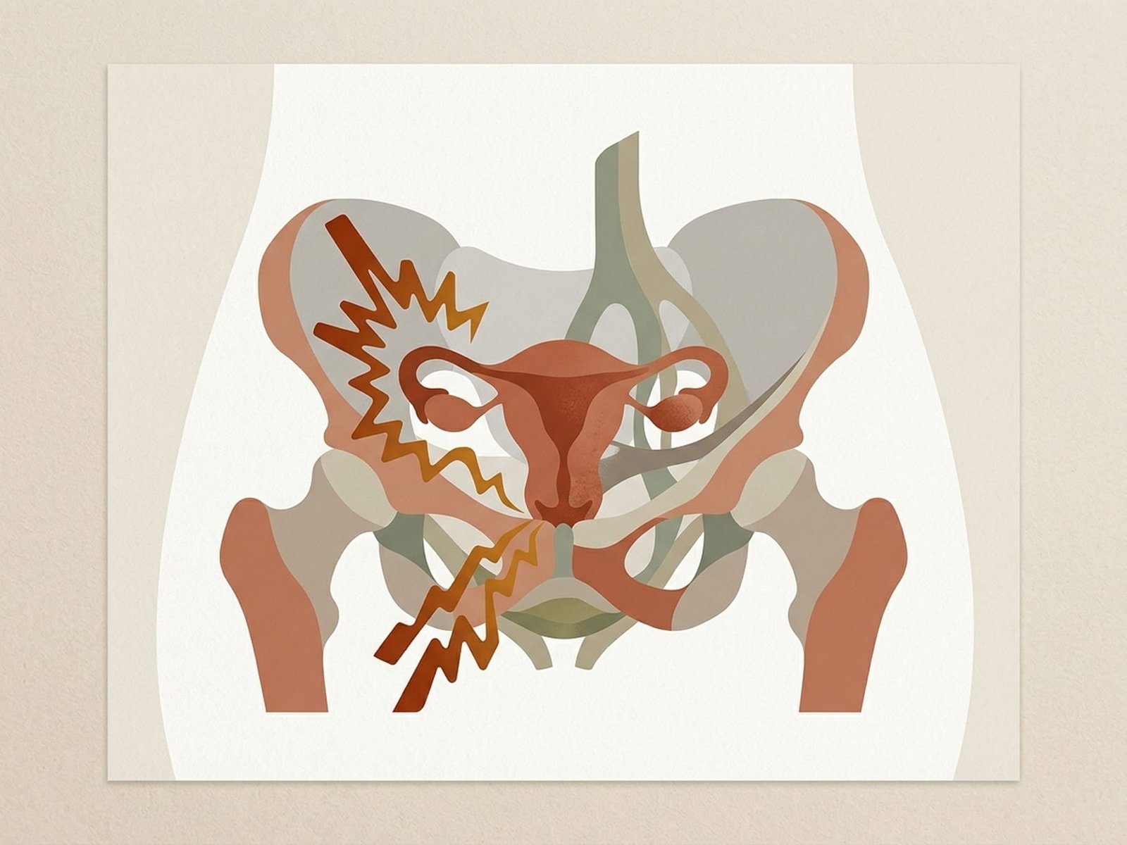
The pelvis contains several layers of muscle that support pelvic organs, contribute to continence and sexual function, and help stabilize the trunk and hip. Trigger points can develop in many of these muscles, but the following are among the most clinically significant contributors to myofascial chronic pelvic pain.
Chronic Pelvic Pain
The levator ani complex — comprising the pubococcygeus, puborectalis, and iliococcygeus — forms the muscular floor of the pelvis. Trigger points in these muscles can produce deep, aching pelvic pain that patients describe as pressure or heaviness in the perineum, vagina, or rectum. Pelvic floor trigger points are an important and often under-recognized contributor to chronic pelvic pain and may overlap with diagnostic labels such as interstitial cystitis or chronic prostatitis/chronic pelvic pain syndrome.
A deep external hip rotator that lines the inner wall of the pelvis before exiting through the lesser sciatic foramen. Trigger points in the obturator internus can refer pain to the posterior pelvis, coccyx, and posterior thigh. Because of its anatomical proximity to the pudendal nerve, a highly irritable or hypertonic obturator internus may also contribute to pudendal-neuralgia-like symptoms including vulvar or scrotal burning, urethral discomfort, and sitting pain.
The piriformis is a deep hip external rotator that lies close to the sciatic nerve. In the context of pelvic pain, piriformis trigger points can refer pain into the deep buttock, sacroiliac region, and posterior pelvis. In some patients, piriformis hypertonicity may also contribute indirectly to pudendal-region symptoms or to sitting pain that is mistakenly attributed only to anorectal causes.
The lower fibers of the rectus abdominis, particularly near the pubic attachment, are a frequently overlooked source of referred pelvic pain. Trigger points in this region may refer pain to the suprapubic area, inguinal region, and across the lower pelvis in a horizontal band. Lower rectus abdominis trigger points can overlap with bladder pain, menstrual cramping, or inguinal pain and are commonly activated by abdominal surgery, prolonged core bracing, or postpartum abdominal wall changes.
The psoas major and iliacus together form the primary hip flexor complex. Trigger points in the iliopsoas may refer pain deep into the anterior pelvis, groin, and medial thigh. Patients with iliopsoas involvement often report difficulty standing fully upright, pain that worsens with prolonged sitting, and a sensation of deep abdominal or pelvic cramping. Iliopsoas contribution is frequently missed in pelvic pain evaluations because the muscle is difficult to palpate externally.
The largest of the adductor group, the adductor magnus has both adductor and hamstring portions. Trigger points may refer pain to the medial thigh, deep groin, and into the pelvis. The hamstring portion can also refer pain toward the ischial tuberosity region, overlapping with ischial bursitis or proximal hamstring complaints. Adductor magnus trigger points are commonly aggravated by prolonged hip adduction, horseback riding, or pelvic asymmetry.
The coccygeus muscle lies posterior to the levator ani and attaches from the ischial spine to the sacrum and coccyx. Trigger points can produce localized coccygeal pain (coccydynia) with referral to the posterior pelvic floor and lower sacrum. Patients often have difficulty sitting on hard surfaces and may shift weight constantly. Coccygeus trigger points are commonly activated by falls onto the buttocks, prolonged sitting, or childbirth-related strain.
The lower fibers of the internal oblique insert into the inguinal ligament and conjoint tendon. Trigger points in this region may refer pain to the inguinal canal, lower abdomen, and suprapubic area. These symptoms can overlap quite closely with inguinal hernia or abdominal wall pain syndromes. Lower internal oblique trigger points are commonly activated by repetitive twisting, heavy lifting, and chronic coughing.
Pelvic Floor (Levator Ani)
Deep muscular sling producing perineal pressure, vaginal or rectal ache, and sitting pain — a major myofascial contributor to chronic pelvic pain.
Obturator Internus
Deep hip rotator whose proximity to the pudendal nerve can help explain burning, urethral pain, and dyspareunia-like symptoms.
Iliopsoas Complex
Hip flexor trigger points can create deep anterior pelvic cramping, groin pain, and difficulty with prolonged sitting or standing upright.
Lower Rectus Abdominis
Abdominal wall trigger points referring suprapubic pain in a horizontal band — often overlapping with bladder pain or menstrual cramping.
Referral Patterns
Pelvic referral patterns help trace symptoms back to a possible myofascial source. Pelvic trigger point referral can overlap so closely with visceral organ pain that patients often undergo prolonged urological, gynecological, or gastroenterological investigation before the muscular component is considered.
Pelvic Floor (Levator Ani)
Obturator Internus
Iliopsoas
Rectus Abdominis (Lower)
Coccygeus
Self-Assessment
The following clinical tests and self-assessment maneuvers can help identify myofascial trigger point contributors to chronic pelvic pain. Because internal pelvic examination is specialized, many of these assessments are best performed by a trained pelvic floor physical therapist or clinician, but several external tests can still support early screening.
External Pelvic Floor Palpation
Obturator Internus Stretch Test
Lower Rectus Abdominis Palpation
Thomas Test (Modified for Pelvic Pain)
Seated Provocation Test
Treatment Pathways
Effective management of myofascial pelvic pain often requires a specialized, multimodal approach that differs from standard generic chronic pain protocols. A key principle is that many pelvic pain patients need pelvic floor relaxation and down-training rather than strengthening alone. Some patients have been worsened by unsupervised Kegel programs when the actual problem was hypertonicity rather than weakness.

Treatment Pathways
Mechanism DiagramSelf-Care
- Pelvic floor relaxation breathing — diaphragmatic breathing with conscious pelvic floor release on exhalation, 10 minutes twice daily
- Adductor and hip rotator stretching — butterfly stretch, piriformis stretch, and deep squat hold for 30 seconds each, 3 repetitions
- Warm sitz baths (15-20 minutes) to promote pelvic floor muscle relaxation and reduce trigger point irritability
- Avoid prolonged sitting — use a standing desk or cushioned seat, take standing breaks every 30 minutes
- Self-release with a tennis ball or pelvic floor wand for external perineal and internal pelvic floor trigger points, when appropriate instruction and safety guidance have been provided
Manual Therapy
- Internal pelvic floor myofascial release by a trained pelvic floor physical therapist — often a key treatment option for levator ani and obturator internus trigger points
- External trigger point release of the lower abdominals, adductors, piriformis, and hip rotators
- Myofascial release of the thoracolumbar fascia and abdominal fascia to address broader continuity patterns when relevant
- Connective tissue or soft-tissue techniques around the sacral and perineal regions when clinically indicated
- Visceral mobilization approaches may be considered by some clinicians in selected cases, though evidence and clinical fit vary
Interventional
- Trigger point dry needling of the obturator internus, piriformis, and selected pelvic floor muscles when performed by appropriately trained practitioners
- Trigger point injection with local anesthetic into accessible pelvic muscles for selected patients
- Pudendal nerve block for diagnostic or therapeutic purposes when pudendal neuralgia overlaps with myofascial pain
- Botulinum toxin injection into pelvic floor muscles may be considered in refractory hypertonic pelvic floor dysfunction, though it is not a universal first-line treatment
Rehabilitation
- Pelvic floor down-training — learning to relax rather than strengthen the pelvic floor, often the opposite of traditional Kegel exercises
- Hip strengthening with emphasis on gluteus medius and external rotators to reduce compensatory pelvic floor overload
- Core stabilization retraining with emphasis on coordination rather than chronic bracing — transversus abdominis and diaphragm integration
- Graded exposure to sitting, sexual activity, and other provocative activities as pain settles
- Cognitive behavioral therapy and pain neuroscience education when chronic pelvic pain is accompanied by fear, guarding, or broader sensitization features
Red Flags — When to See a Doctor
Before attributing pelvic pain to a myofascial source, always screen for the following red flags that may indicate serious underlying pathology requiring urgent evaluation.
Chronic pelvic pain is common, yet a meaningful myofascial component is frequently under-recognized in both men and women.
The pelvic floor muscles, obturator internus, and lower abdominal muscles are among the most clinically important myofascial contributors. Their referral patterns can closely overlap with interstitial cystitis, chronic prostatitis/chronic pelvic pain syndrome, endometriosis, vulvodynia, and abdominal wall pain syndromes.
Pelvic floor myofascial pain often requires a different strategy from standard strengthening-based pelvic rehabilitation. In patients with hypertonic pelvic floor dysfunction, relaxation and down-training are often more appropriate than Kegel exercises.
Internal pelvic floor examination and treatment by a trained pelvic floor physical therapist is often one of the most useful approaches, especially for levator ani and obturator internus involvement.
The obturator internus is a particularly important and often overlooked muscle whose relationship to the pudendal nerve can help explain burning, urethral pain, dyspareunia, and sitting-aggravated pelvic pain.
Always screen for red flags before attributing pelvic pain primarily to a myofascial source — hematuria, post-menopausal bleeding, fever, neurological symptoms, and acute severe onset all warrant urgent evaluation.