Understanding the Clinical Overlap
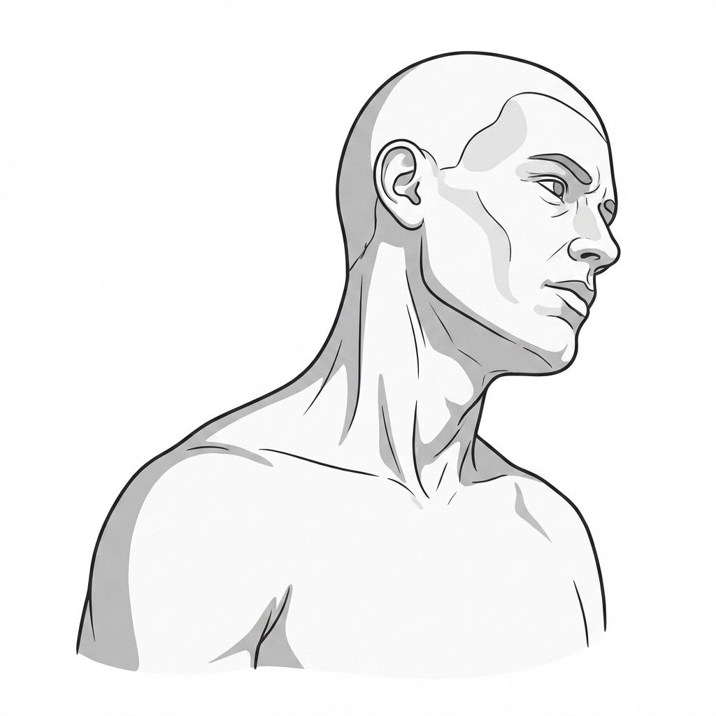
Cervical dystonia (CD), historically referred to as spasmodic torticollis, is a focal dystonia characterized by involuntary muscle contractions in the neck. These contractions cause abnormal movements and postures of the head and neck, most commonly rotational (torticollis), but also lateral tilt (laterocollis), forward flexion (anterocollis), or backward extension (retrocollis). Current pathophysiological models suggest CD originates from dysfunction within the basal ganglia and its sensorimotor network connections. Unlike many other movement disorders, CD is highly associated with pain, with epidemiological data indicating that most patients experience significant cervical discomfort. Differentiating the central motor drive of CD from peripheral musculoskeletal conditions is a critical first step in clinical management.
The relationship between cervical dystonia and myofascial pain syndrome (MPS) is complex and frequently bidirectional. The chronic, involuntary contractions inherent to CD place immense biomechanical strain on both the primary dystonic muscles and the antagonist muscles attempting to stabilize the cervical spine. This sustained metabolic demand and localized ischemia create an ideal environment for the development of secondary myofascial trigger points. Consequently, a patient with CD often presents with a superimposed MPS. The pain experienced by the patient is frequently a composite of the deep, aching cramp of the dystonic spasm and the sharp, referred pain characteristic of active myofascial trigger points in muscles like the upper trapezius, levator scapulae, and splenii.
While myofascial pain syndrome primarily involves peripheral muscle sensitization, cervical dystonia is fundamentally a disorder of central motor control, though the two frequently coexist and amplify one another.
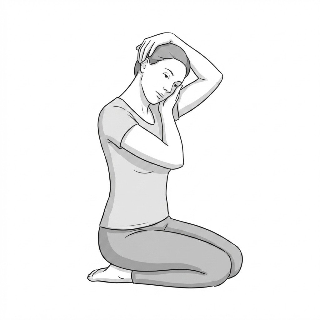
Differentiating primary MPS from CD, or identifying their coexistence, requires careful clinical observation. A hallmark of CD is the "geste antagoniste" or sensory trick, where light tactile stimulation temporarily alleviates the dystonic posturing; this phenomenon is absent in pure MPS. Furthermore, while MPS is characterized by discrete taut bands and localized twitch responses upon palpation, CD often presents with global muscle hypertrophy and continuous or tremulous rigidity. Current clinical consensus suggests that while botulinum toxin remains the first-line treatment for the dystonic component, targeted manual therapy and rehabilitation addressing the secondary myofascial pain can significantly improve overall patient outcomes. Recognizing this dual pathology prevents the common clinical error of treating a central motor disorder solely with peripheral soft tissue techniques.
Key Musculature in Dystonia and MPS
The clinical presentation of cervical dystonia depends on which specific muscles are receiving abnormal central motor drive. Understanding the primary actors versus the secondary compensatory muscles is essential for both diagnosis and targeted intervention.
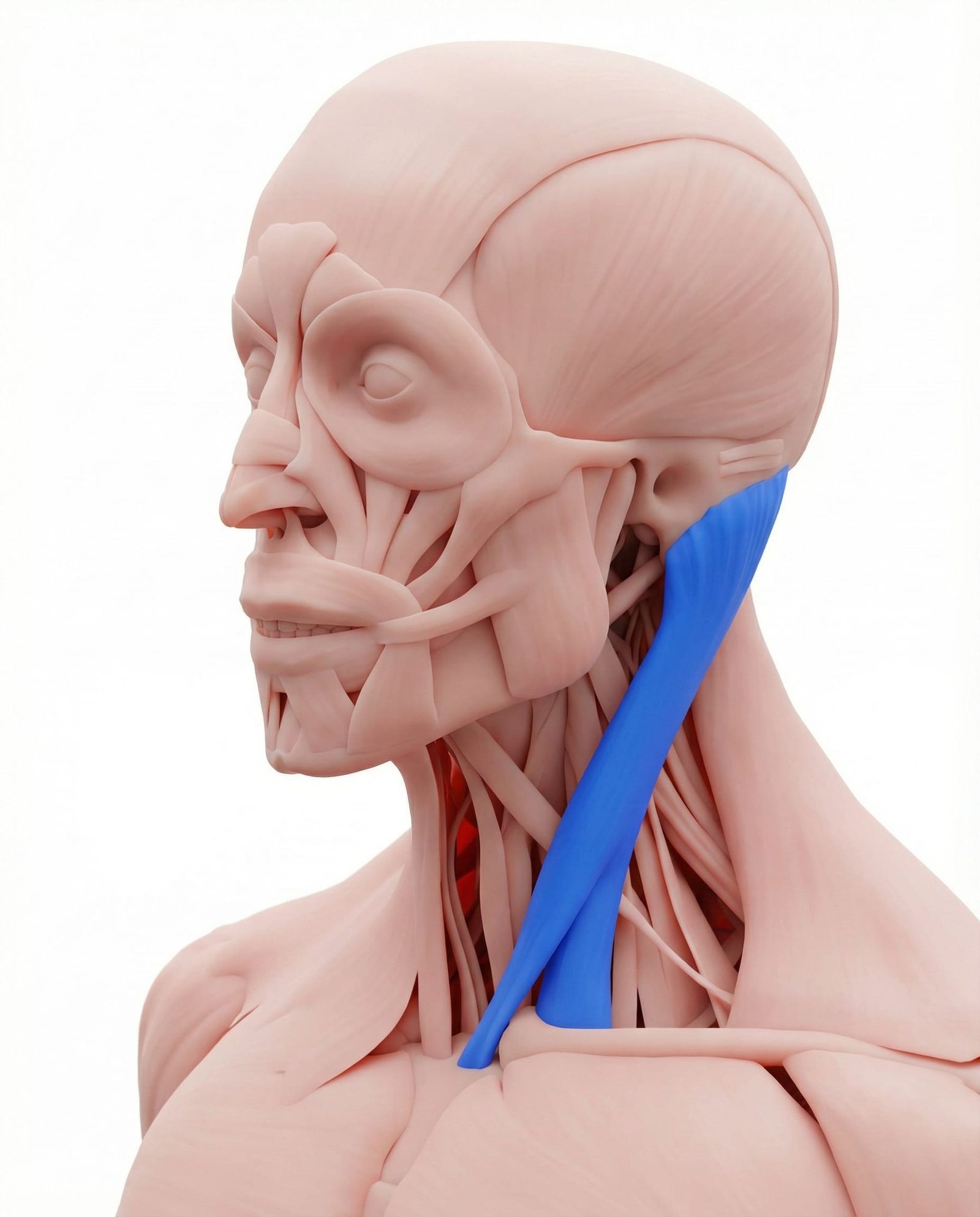
The SCM is frequently the primary driver of rotational torticollis in cervical dystonia, often exhibiting visible hypertrophy due to chronic involuntary contraction. In myofascial pain syndrome (MPS), the SCM develops discrete taut bands and trigger points without the continuous dystonic drive. Differentiating between dystonic contraction and myofascial taut bands in this muscle is critical for appropriate intervention. Coexisting MPS in a dystonic SCM can significantly amplify the patient's pain burden.
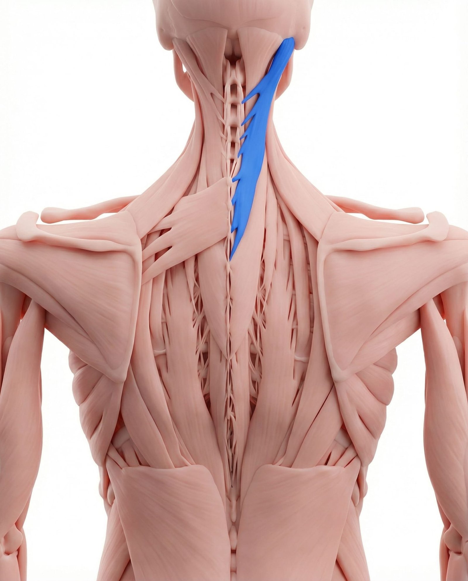
These posterior cervical muscles act as primary ipsilateral rotators and extensors. In cervical dystonia, they frequently act synergistically with the contralateral SCM to produce complex rotational and extension posturing (torticollis and retrocollis). Secondary myofascial trigger points routinely develop in the splenii due to the constant biomechanical strain of opposing the dystonic pull. Clinical assessment often reveals significant tenderness and referred pain originating from these overloaded tissues.
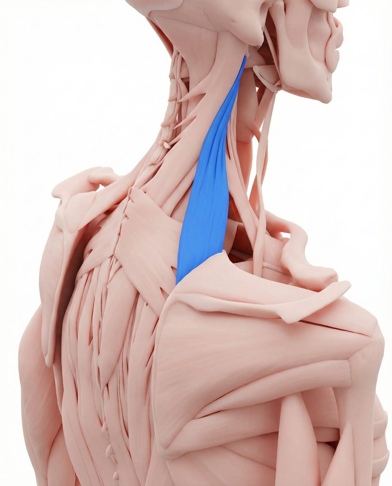
The levator scapulae is commonly involved in laterocollis (side-bending) and retrocollis presentations of cervical dystonia. Because it elevates the scapula and rotates the cervical spine, it is highly susceptible to secondary myofascial overload when compensating for abnormal head postures. Patients frequently report a "stiff neck" sensation that may be primarily myofascial in origin, even if the underlying driver is dystonic. Manual assessment must distinguish between dystonic rigidity and myofascial shortening.
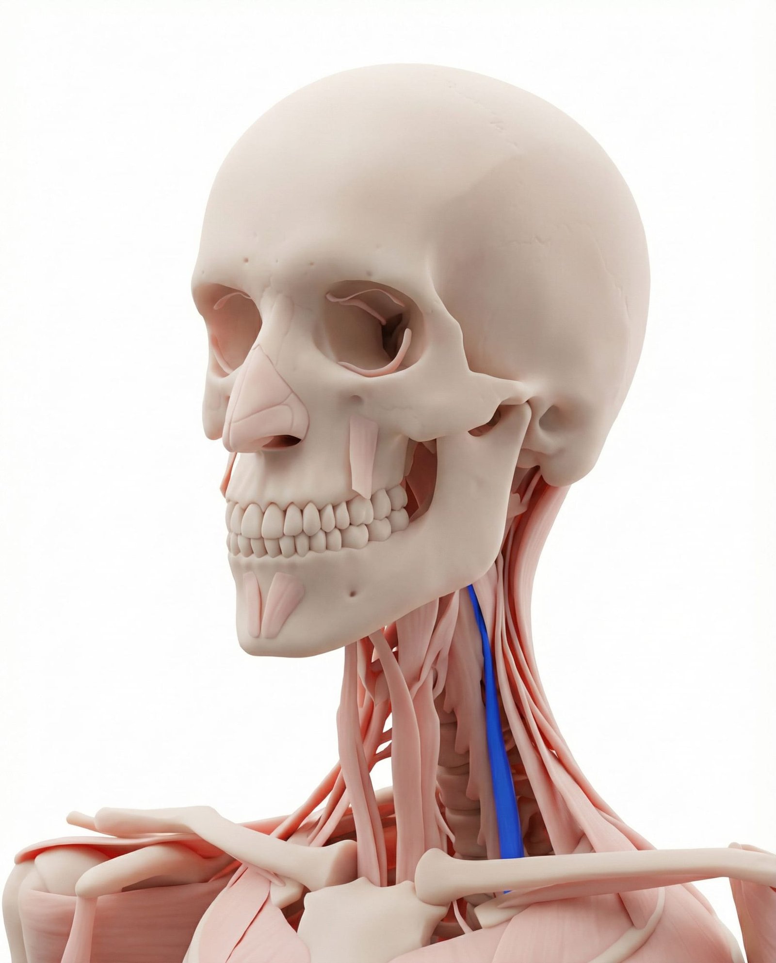
While less commonly the primary drivers of cervical dystonia compared to the SCM or splenii, the scalenes are frequently recruited in laterocollis or anterior shift postures. Chronic dystonic posturing can lead to secondary scalene shortening and subsequent myofascial trigger point formation. This secondary involvement is clinically significant as it may contribute to thoracic outlet-like symptoms or complex regional pain presentations. Careful palpation is required to identify scalene involvement beneath the hypertrophied SCM.
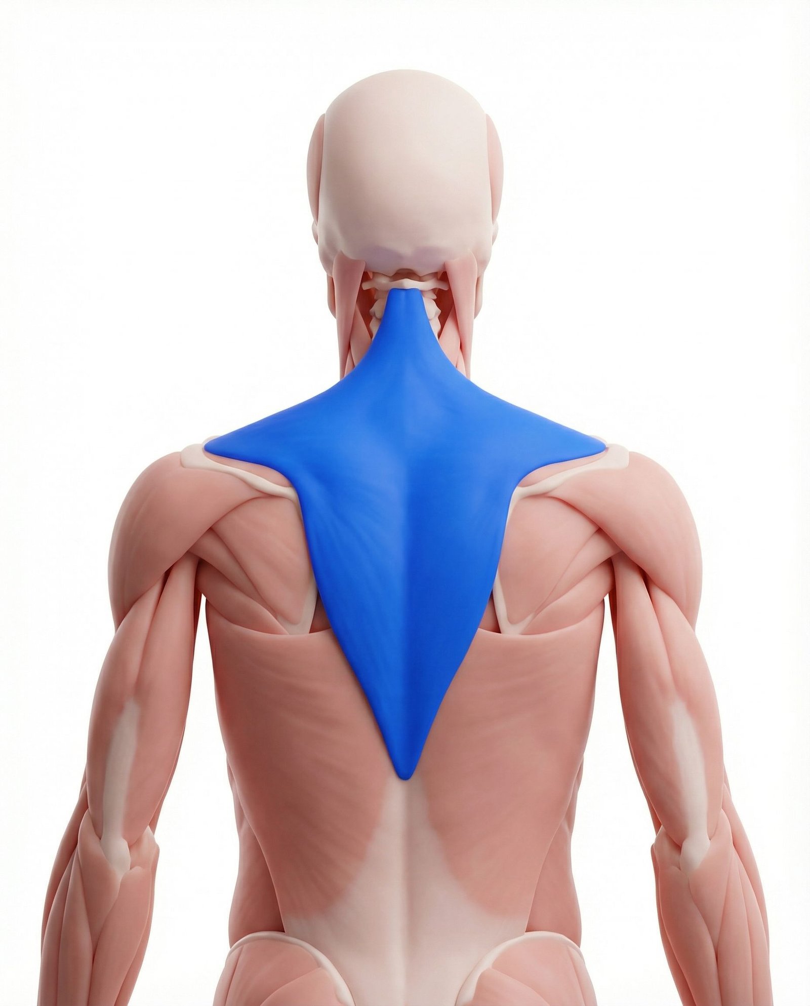
Shoulder elevation is a common compensatory or primary dystonic movement accompanying cervical dystonia. The upper trapezius frequently develops severe myofascial pain syndrome as a secondary consequence of this chronic elevation and altered cervical biomechanics. Treating the myofascial component in the upper trapezius often provides meaningful symptomatic relief, even when the central dystonic drive remains active. It serves as a prime example of the intersection between central motor disorders and peripheral pain syndromes.
Basal Ganglia Dysfunction
Altered sensorimotor integration in the central nervous system leads to continuous, involuntary efferent drive to cervical musculature, causing painful cramping.
Secondary Myofascial Sensitization
Sustained contraction leads to localized ischemia, ATP depletion, and the formation of active myofascial trigger points in both dystonic and compensatory muscles.
Biomechanical Overload
Abnormal head postures alter the normal axis of the cervical spine, placing severe mechanical stress on facet joints, ligaments, and antagonist muscle groups.
Central Sensitization
Chronic nociceptive input from sustained muscle contraction and trigger points can lead to upregulation of the central nervous system, lowering pain thresholds globally.
Pain Referral Patterns
Pain in cervical dystonia is rarely confined to the actively contracting muscles. Secondary myofascial trigger points generate predictable referral patterns that can mimic primary headache disorders or cervical radiculopathy.
Sternocleidomastoid (SCM)
Splenius Cervicis
Upper Trapezius
Levator Scapulae
Clinical Differentiation Tests
Clinical differentiation between cervical dystonia and primary myofascial pain relies on specific observational and physical examination techniques. These assessments help distinguish central motor dysfunction from peripheral tissue sensitization.
Sensory Trick (Geste Antagoniste) Evaluation
Active Range of Motion (AROM) and Null Point Assessment
Palpation for Hypertrophy vs. Taut Bands
Dystonic Tremor Observation
Comprehensive Management Pathways
Management of cervical dystonia with comorbid myofascial pain requires a multimodal approach. Current guidelines generally recommend medical intervention for the central motor drive, paired with rehabilitation for the peripheral tissue adaptations.
Self-Care
- Identification and utilization of effective sensory tricks (geste antagoniste)
- Ergonomic modifications to support the head and reduce sustained muscular effort
- Stress management and relaxation techniques to modulate central nervous system arousal
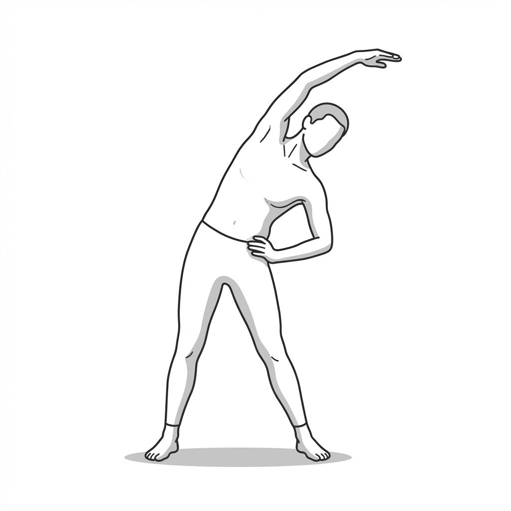
- Gentle, pain-free active range of motion exercises within tolerated limits
- Application of localized heat to manage secondary muscle aching and stiffness
Manual Therapy
- Targeted trigger point release for secondary myofascial pain in compensatory muscles
- Gentle soft tissue mobilization to maintain tissue extensibility
- Avoidance of forceful manipulation or aggressive stretching of dystonic muscles
- Myofascial release techniques focused on the upper trapezius and levator scapulae
- Proprioceptive neuromuscular facilitation (PNF) adapted for dystonia tolerance
Interventional
- Botulinum toxin injections (generally considered the first-line medical treatment)
- Electromyography (EMG) or ultrasound guidance to ensure precise toxin placement
- Trigger point injections for severe, localized secondary myofascial pain
- Oral pharmacological management (e.g., anticholinergics, muscle relaxants) as adjuncts
- Neurology consultation for consideration of deep brain stimulation (DBS) in refractory cases
Rehabilitation
- Neuromuscular re-education to improve motor control and proprioception
- Biofeedback therapy to assist in recognizing and reducing involuntary contractions
- Postural adaptation strategies to minimize biomechanical strain
- Strengthening of antagonist muscles to provide better cervical stability
- Coordination with neurology for optimal timing of therapy post-botulinum toxin injection
Red Flags — When to See a Doctor
While cervical dystonia is typically a chronic, idiopathic condition, acute onset or the presence of specific neurological signs may indicate a severe underlying pathology requiring immediate medical investigation.
Clinical Pearls
Cervical dystonia is a central nervous system disorder of motor control, whereas myofascial pain syndrome is a peripheral neuromuscular condition, though the two frequently coexist.
The presence of a sensory trick (geste antagoniste) is highly specific to dystonia and is a critical clinical feature for differentiating it from pure myofascial or structural torticollis.
Secondary myofascial trigger points routinely develop in muscles compensating for dystonic posturing, significantly contributing to the patient's overall pain burden.
Current neurological guidelines generally recommend botulinum toxin injections as the first-line evidence-based intervention for primary cervical dystonia.
Physical therapy for cervical dystonia should prioritize neuromuscular re-education and management of secondary myofascial pain rather than aggressive stretching of dystonic muscles.
Treating the secondary myofascial component can provide meaningful symptomatic relief and improve quality of life, even if the underlying dystonic drive remains active.
Movement therapy
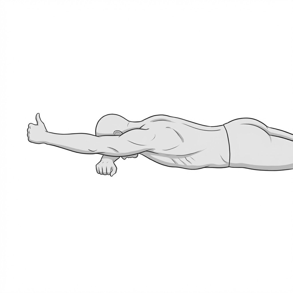
Exercises for Cervical Dystonia
These exercises work with the aberrant motor drive of dystonia rather than against it — antagonistic isometrics and sensory-trick training help recalibrate cervical motor output, while gentle suboccipital release reduces the secondary compression that amplifies the sustained posture. Start with 2–3 and progress as tolerated.