Understanding Breathing Pattern in MPS
Breathing Mechanics
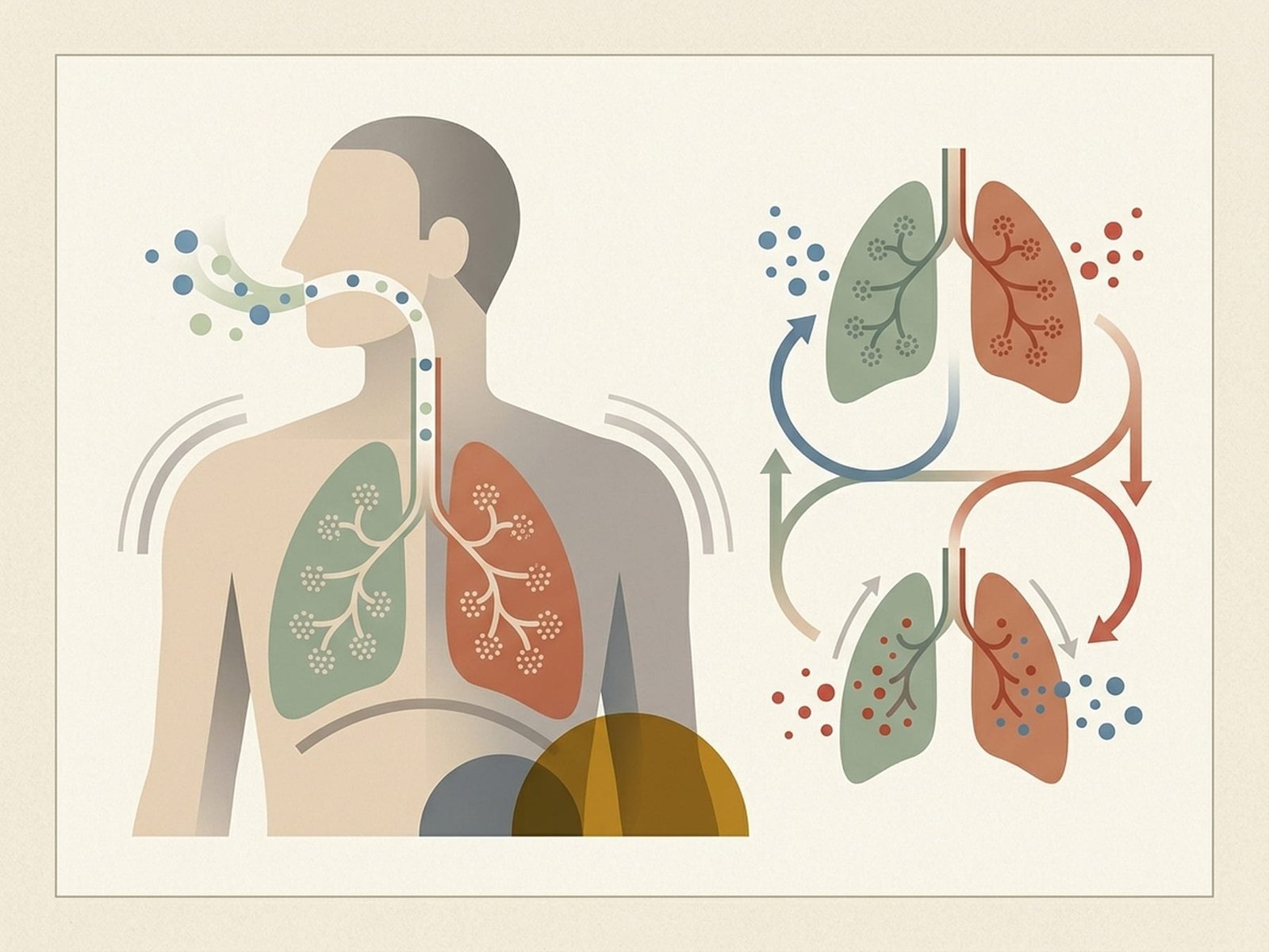
Diaphragm vs. Upper Chest Pattern OverviewBreathing is one of the few bodily functions that is both automatic and modifiable. In some patients with chronic myofascial pain — especially in the neck, shoulder girdle, upper thorax, or chest wall — breathing pattern appears to be part of the perpetuating picture. A common pattern is upper-chest or accessory-muscle-dominant breathing, in which the scalenes, SCM, and upper chest muscles work harder than they need to during routine breathing.
This can matter mechanically because muscles used repeatedly as breathing assistants may also become overloaded as postural or movement muscles. If the diaphragm and lower rib cage contribute less effectively, accessory muscles may stay busy throughout the day. Over time, this may help explain why some cervico-thoracic trigger points keep recurring unless breathing mechanics are also addressed.
Breathing pattern is not only mechanical. It also interacts with stress physiology, autonomic arousal, pain vigilance, and movement guarding. Some people breathe faster, higher, or more shallowly when in pain or under stress, and that pattern can become habitual. In those cases, breathing retraining may help by reducing unnecessary effort, improving tolerance of breathing sensations, and reducing accessory-muscle load.
Breathing retraining is appealing because it is inexpensive, accessible, and relevant to several common perpetuating factors at once. At the same time, it should not be overclaimed. Not every patient with myofascial pain has meaningful breathing dysfunction, and not every symptom pattern is primarily respiratory in origin. The most useful role of this page is to help identify when breathing deserves attention as part of a broader treatment plan.
Respiratory Muscles & MPS
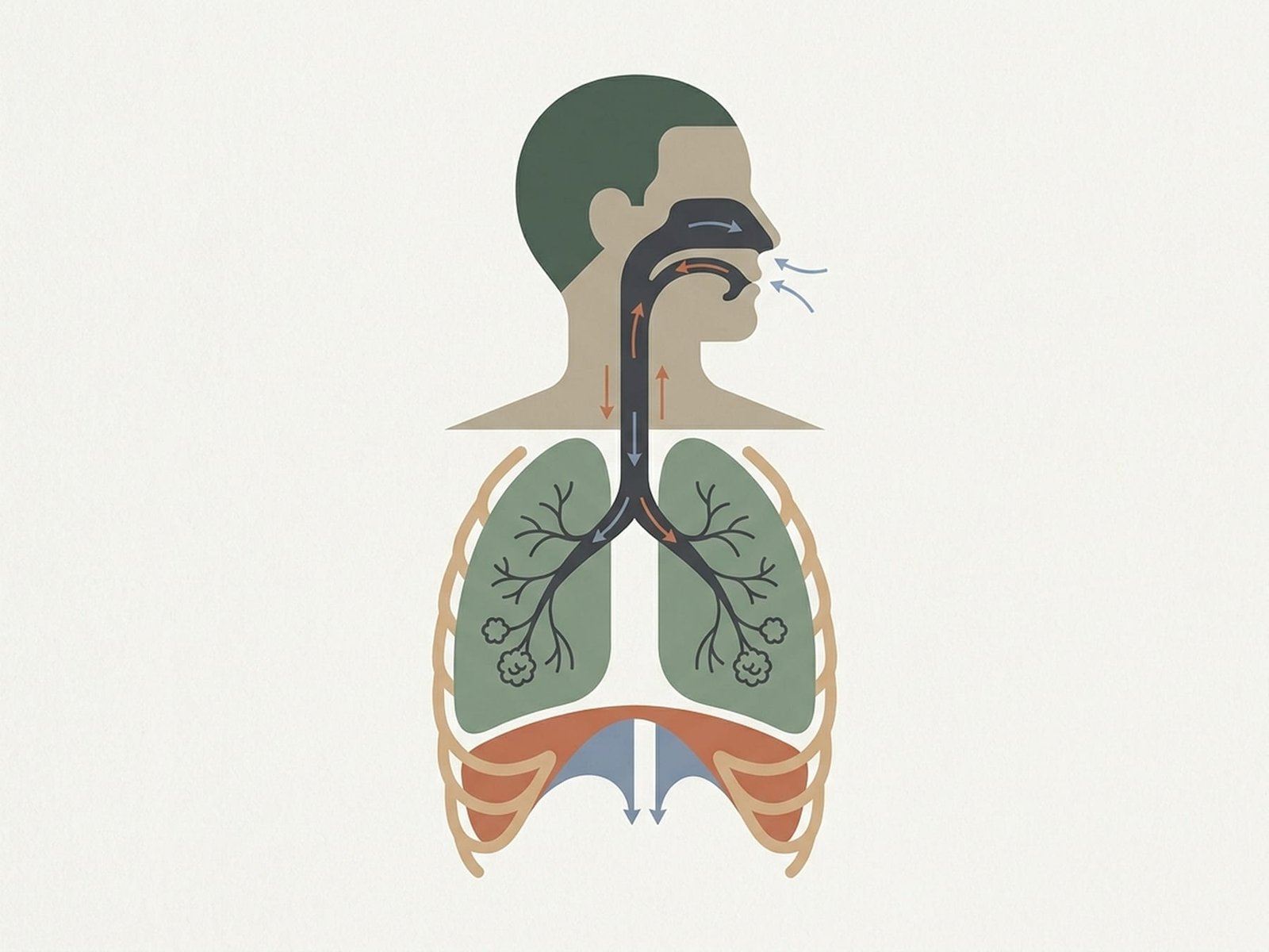
Several muscle groups sit at the intersection of breathing mechanics and myofascial pain. Understanding their roles helps explain why a person can have both breathing-pattern dysfunction and persistent trigger point activity in the neck, shoulder girdle, chest wall, or upper back.
Respiratory Muscles
Scalenes (Anterior, Middle, Posterior)
Sternocleidomastoid (SCM)
Pectoralis Minor
Diaphragm
Intercostal Muscles
Upper Chest Breathing
Accessory muscles work more than necessary, which may increase load in the neck and upper thorax over time.
Diaphragm-Led Breathing
The usual target pattern in retraining — quieter lower-rib and abdominal contribution with less unnecessary upper-chest effort.
Breath Holding
Common during pain, effort, or stress; may increase chest wall and accessory-muscle tension.
Paradoxical Breathing
May range from a benign habitual pattern to a sign of more significant respiratory dysfunction depending on context.
Referral Patterns
Respiratory and accessory breathing muscles can produce symptom patterns that overlap with many other musculoskeletal and non-musculoskeletal conditions. Scalene-related symptoms may extend into the chest, shoulder, arm, or hand. SCM-related symptoms may be experienced in the head, face, or jaw. Pectoralis minor and intercostal symptoms can contribute to chest wall pain and breathing discomfort. None of these patterns are specific enough to stand alone as a diagnosis, which is why context and differential assessment remain important.
One useful clinical clue is that respiratory-muscle-related symptoms sometimes change with breathing depth, rib cage movement, thoracic posture, or breathing pattern. That does not prove a breathing-driven cause, but it can justify looking more carefully at breathing mechanics as part of the assessment.
When breathing relies too heavily on the neck and upper chest, those muscles may eventually become part of the pain problem rather than just part of the breathing pattern.
Scalene Referral
May involve chest, shoulder, arm, or hand symptoms
SCM Referral
May overlap with head, face, jaw, or dizziness-related complaints
Chest Wall Involvement
Intercostals and pectoral region may influence breathing-related pain
Breathing Assessment Tests
These assessments can help identify whether breathing pattern, breathing rate, chest wall mobility, or breath-hold intolerance might be relevant. They are best used as part of a broader clinical picture rather than as stand-alone tests.
Hi-Lo Hand Test
Breathing Rate at Rest
BOLT Score
Thoracic Mobility Assessment
Treatment Pathways
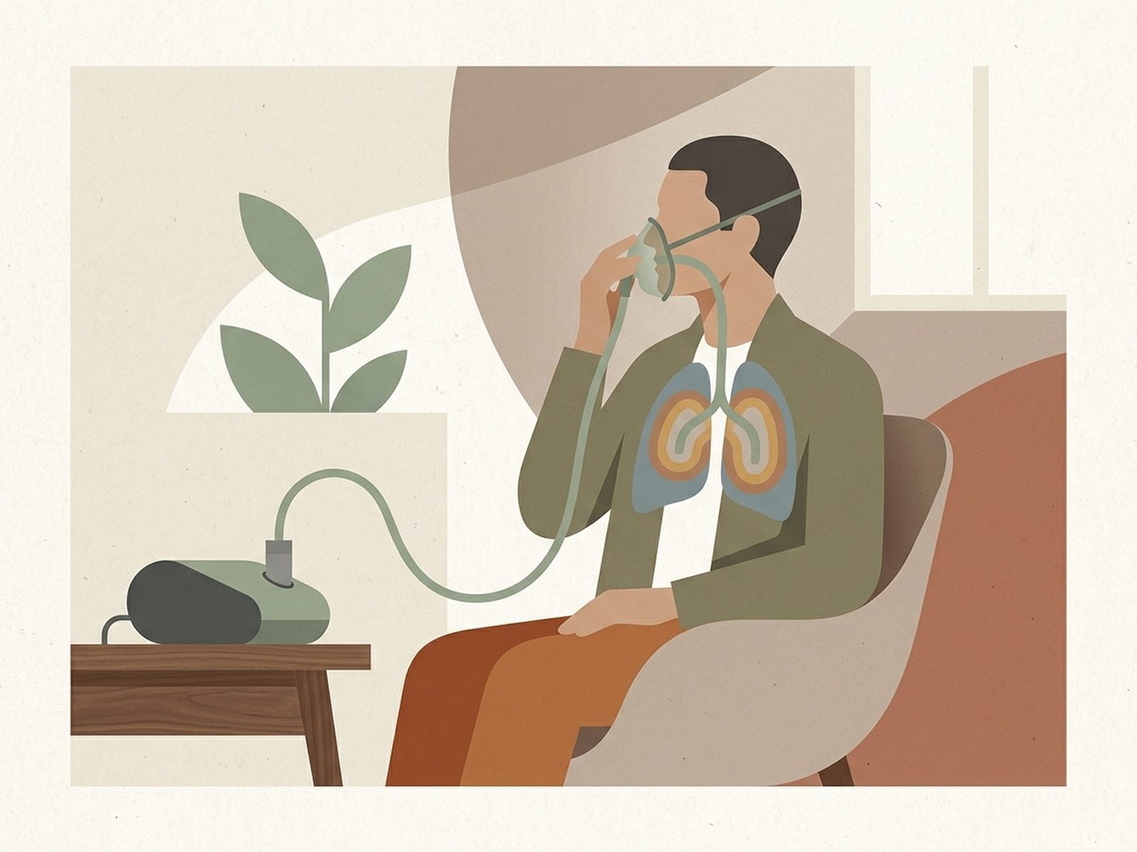
Breathing retraining usually works best as a gradual progression rather than a forceful correction. In practice, many patients do better when they first learn an easier pattern, then slowly integrate it into posture, movement, and everyday activities.
Breathing Retraining Protocols
Progression DiagramDiaphragmatic Breathing Practice
For many patients, the most useful starting point is practicing slower, lower-rib and abdominal breathing in a comfortable position. The goal is not perfect technique immediately, but gradually reducing unnecessary accessory-muscle overuse.
- Begin supine or semi-reclined with knees bent if that feels easiest
- Inhale gently through the nose, allowing the abdomen and lower rib cage to expand without forcing the chest upward
- Use a slightly longer exhale if comfortable; some patients find this calming and easier to sustain
- Practice briefly but consistently rather than forcing long sessions early on
- Avoid exaggerated “belly pushing” or breath-holding — the aim is quieter, easier breathing rather than performance
Breathing Rate and CO2 Tolerance Work
Some patients with overbreathing, anxiety-related breathing dysregulation, or strong accessory-muscle dominance benefit from gradual work on slower breathing and better tolerance of normal respiratory sensations. Buteyko-style strategies are one example of this approach, though they should be presented as one model rather than the only correct method.
- Encourage nasal breathing when practical and comfortable, especially during quiet breathing
- Use brief breath-awareness drills or light pause-based work only if they are well tolerated
- Slowing breathing should feel manageable, not forced or alarming
- Track symptoms such as dizziness, air hunger, or panic rather than pushing for arbitrary numbers
- If a BOLT-style score is used, treat it as a rough internal tracking measure rather than a universal target
Postural Breathing Integration
Breathing pattern often interacts with posture, rib cage position, thoracic mobility, and shoulder girdle mechanics. In some patients, retraining works better when breathing practice is paired with posture and movement work rather than treated as an isolated exercise.
- Practice easier lower-rib breathing in upright postures, not only lying down
- Use thoracic extension or rib mobility work when stiffness clearly limits comfortable breathing
- Some people respond well to “360-degree” or lateral rib expansion cues rather than only abdominal cues
- If scalenes or pectoralis minor are highly irritable, symptom reduction in those areas may make retraining easier
- Gradually bring breathing awareness into daily activities rather than limiting it to drills only
Progressive Breathing Training
Lasting change usually comes from gradual integration rather than a quick correction. Breathing retraining tends to work best when it progresses from simple practice to real-life use over time.
- Start with a position that feels easiest and least threatening
- Progress to seated and standing practice when the pattern becomes more comfortable
- Later, integrate breathing during light movement such as walking or easy exercise
- Advance gradually based on tolerance rather than a rigid timeline
- Expect breathing pattern change to take repetition and consistency rather than immediate perfection
Red Flags — When to See a Doctor
Breathing retraining is often safe, but some respiratory or chest-related symptoms require medical evaluation before self-directed exercises are emphasized. The following features deserve caution or escalation.
Breathing pattern can be an important perpetuating factor in some cervico-thoracic and chest-wall myofascial pain presentations.
When accessory muscles such as the scalenes and SCM are recruited excessively during routine breathing, they may become overworked and irritable over time.
Improving diaphragmatic contribution, rib cage movement, and breathing efficiency may help reduce load on accessory muscles in selected patients.
Breathing retraining is low-cost and often practical, but it should be treated as one supportive part of care rather than a universal cure.
Progress is usually better judged by comfort, function, reduced overuse, and easier breathing rather than by rigid numerical targets alone.