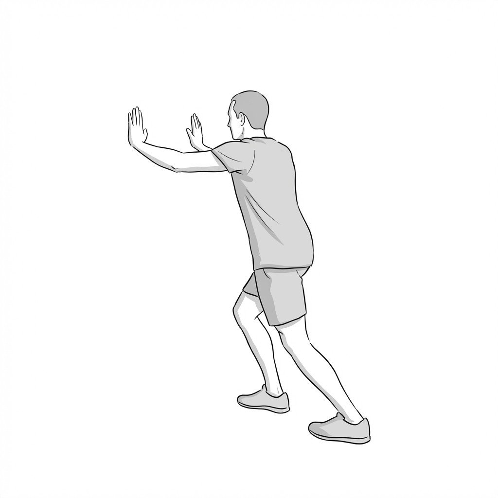
Understanding the Condition
Achilles tendinopathy is a common clinical condition characterized by activity-related pain, localized tendon tenderness, and structural changes within the tendon. It represents a failed healing response to repetitive microtrauma and overload, rather than a primarily inflammatory process. Its prevalence is high in active individuals, particularly runners, but it also affects sedentary populations. Differentiating true tendinopathy from pain referred from myofascial trigger points in the calf muscles is a critical diagnostic challenge, as the treatment approaches differ and the two conditions frequently coexist.
The connection between Achilles tendinopathy and myofascial pain is both anatomical and functional. The gastrocnemius and soleus muscles converge to form the Achilles tendon, and any dysfunction—such as tightness, weakness, or the presence of trigger points—can alter the load distribution and increase strain on the tendon. Furthermore, trigger points in the soleus muscle are well-documented to refer pain directly to the heel and Achilles insertion, perfectly mimicking the symptoms of insertional tendinopathy. This overlap can lead to misdiagnosis or incomplete treatment if the muscular component is not addressed.
Effective management hinges on an accurate diagnosis: are we treating a dysfunctional tendon, dysfunctional muscles, or both?
A thorough clinical evaluation is key to distinguishing between these conditions. The diagnostic process focuses on a detailed history of symptom behavior (e.g., pain with initial steps, "warm-up" phenomenon), specific load tests like the single-leg heel rise, and careful palpation. The clinician aims to identify if the pain originates from the tendon itself, from referred muscular sources, or a combination. Often, successful management requires a dual approach: a structured loading program to restore the tendon's capacity and manual therapy to address the related myofascial dysfunction.
Tendinopathy vs Myofascial Mimic
Pain behavior
Pain location
Palpation finding
First-line treatment
Key Anatomy & Myofascial Contributors
The Achilles tendon is the largest and strongest tendon in the body, formed by the confluence of the gastrocnemius and soleus muscles. Understanding the relationship between these muscles and the tendon is fundamental to diagnosing and treating posterior heel pain.
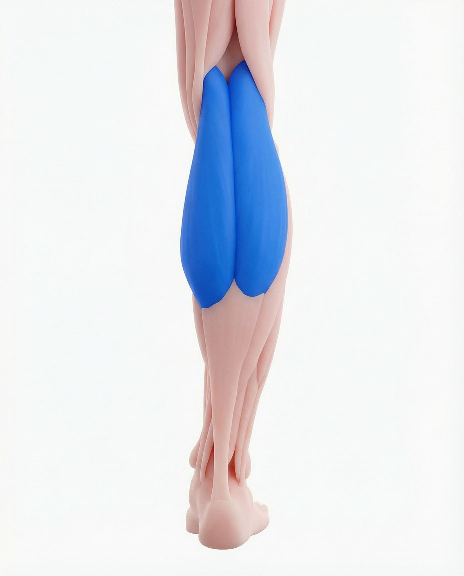
The most superficial calf muscle, the gastrocnemius has two heads (medial and lateral) and crosses both the knee and ankle joints. Due to its biarticular nature and high proportion of fast-twitch fibers, it is susceptible to strain and the development of myofascial trigger points. These trigger points, particularly in the medial head, commonly refer pain distally into the calf, Achilles tendon, and occasionally the plantar arch.
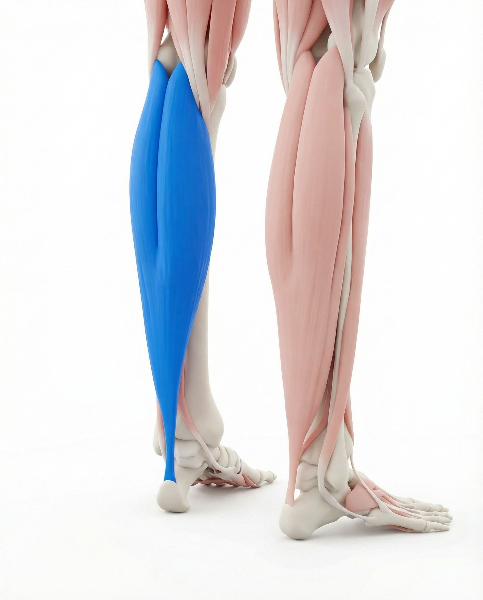
Located deep to the gastrocnemius, the soleus is a powerful, slow-twitch dominant muscle essential for posture and ambulation. It forms the bulk of the Achilles tendon along with the gastrocnemius. Soleus trigger points are a frequent and often overlooked source of "Achilles" pain, with referral patterns that concentrate precisely over the tendon and its insertion on the calcaneus, closely mimicking insertional tendinopathy.
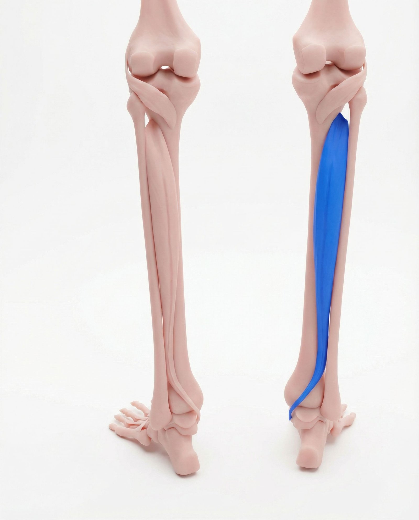
As the deepest muscle in the posterior compartment, the tibialis posterior plays a critical role in supporting the medial longitudinal arch. Dysfunction or myofascial restriction in this muscle can alter foot mechanics, leading to excessive pronation and increased eccentric load on the Achilles tendon. Its trigger points can refer pain directly to the Achilles region, complicating the diagnostic picture.
These muscles of the lateral compartment are primary everters of the foot and dynamically stabilize the ankle. Myofascial trigger points within the peroneals can refer pain to the lateral ankle and heel. This pattern can be confused with the lateral aspect of insertional Achilles tendinopathy or pain from the sural nerve.
The FHL muscle and its tendon run deep and just medial to the Achilles tendon. Tendinopathy or tenosynovitis of the FHL ("dancer's tendonitis") can present with posterior ankle pain that is difficult to distinguish from mid-portion Achilles tendinopathy. Palpation and resisted great toe flexion can help differentiate, but the conditions can also coexist.
Tendon Pathology
Pain originating from disorganized collagen, neovascularization, and neurochemical changes within the tendon substance itself (i.e., true tendinopathy).
Myofascial Referral
Pain referred from active trigger points in the gastrocnemius, soleus, or other deep posterior compartment muscles, which can mimic tendinopathy.
Neurogenic Component
Irritation or entrapment of the sural nerve or tibial nerve can cause pain, tingling, or numbness in the posterior ankle and heel region.
Biomechanical Overload
Pain driven by excessive or abnormal forces on the tendon due to factors like poor foot mechanics (e.g., overpronation), training errors, or deficits elsewhere in the kinetic chain.
Common Myofascial Referral Patterns
Myofascial trigger points are hyperirritable nodules within taut bands of skeletal muscle. When active, they can refer pain to distant sites in predictable patterns. For Achilles pain, the primary muscles of interest are the soleus and gastrocnemius.
Gastrocnemius
Soleus
Tibialis Posterior
Clinical Self-Assessment Tests
These clinical tests are used to systematically assess the structures around the posterior ankle. They help reproduce symptoms in a controlled way to differentiate between tendon pathology and other potential sources of pain. They should be performed gently and within pain tolerance.
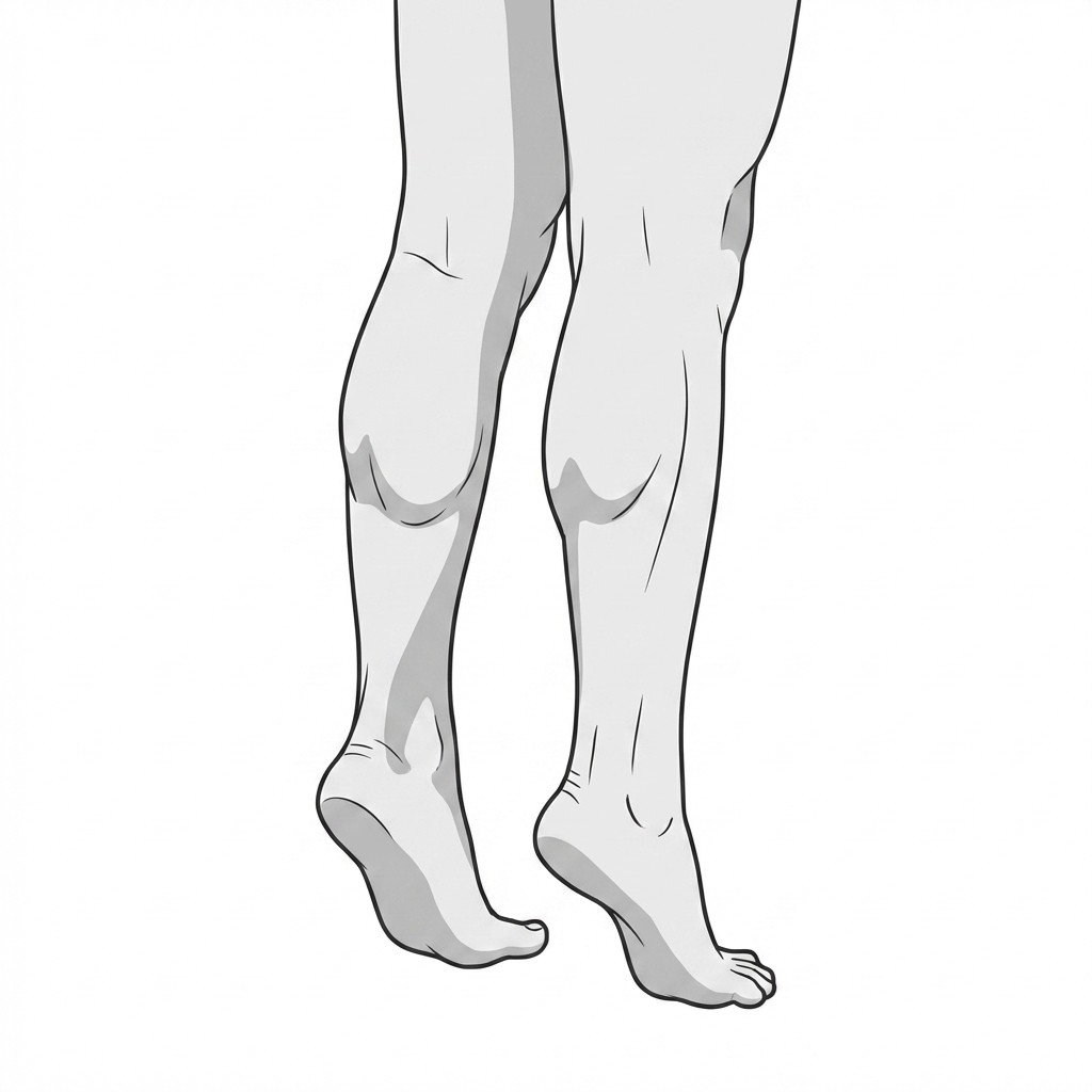
Single-Leg Heel Rise
Painful Arc Sign
Royal London Hospital Test
Palpation for Tendon Thickening
Calf Muscle Palpation
Evidence-Based Treatment Pathways
Modern management of Achilles tendinopathy is centered on progressive load adaptation. The goal is to stimulate tendon remodeling and increase its capacity to handle stress. Adjunctive therapies can be useful for pain modulation and addressing contributing factors like myofascial restrictions.
Rehabilitation
- Progressive tendon loading (cornerstone of treatment)
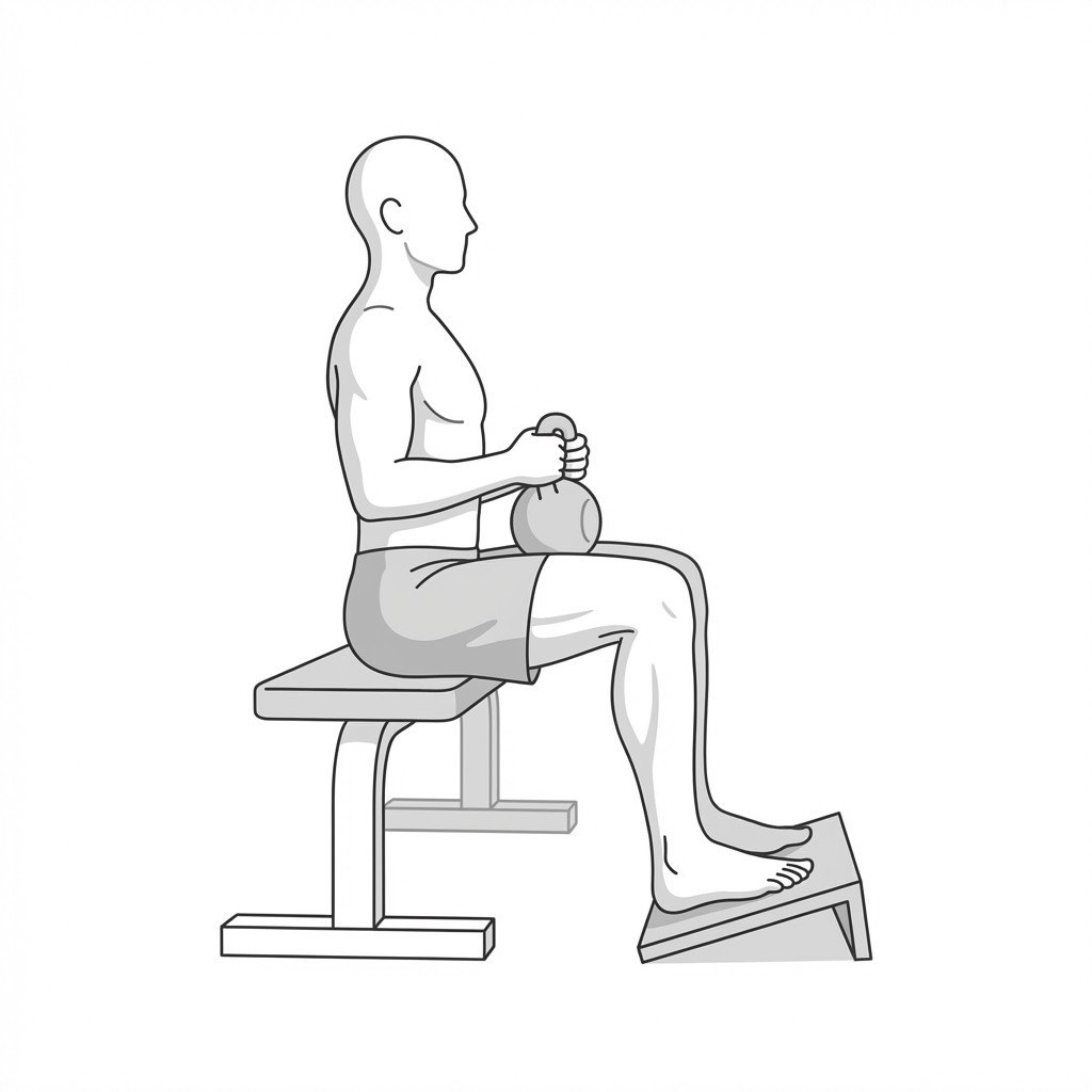
- Heavy slow resistance (HSR) protocols
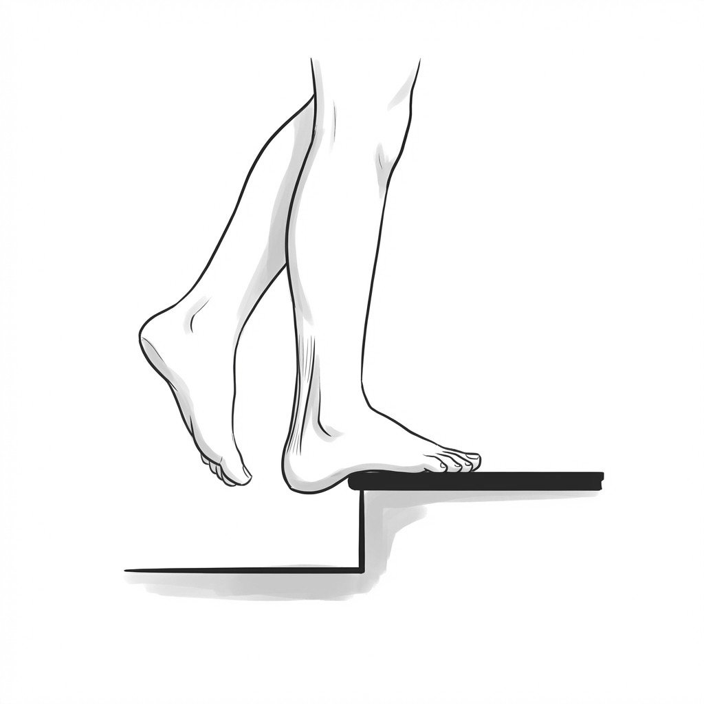
- Eccentric loading exercises (e.g., Alfredson protocol)
- Isometric loading for pain modulation
- Biomechanical assessment and correction (e.g., foot orthoses)
- Kinetic chain strengthening (hip and core)
Manual Therapy
- Soft tissue mobilization for gastrocnemius and soleus
- Trigger point dry needling for calf muscles
- Instrument-assisted soft tissue mobilization (IASTM)
- Joint mobilization for ankle, subtalar, and midfoot joints
- Cross-friction massage over the tendon (use with caution)
Self-Care
- Activity modification and load management
- Pain monitoring during and after activity
- Appropriate footwear with adequate cushioning/support
- Heel lifts (short-term use to unload the tendon)
- Avoiding aggressive static stretching of the painful tendon
- Gentle calf foam rolling or massage
Interventional
- Diagnostic musculoskeletal ultrasound
- Ultrasound-guided peritendinous injections (e.g., high-volume injection)
- Platelet-rich plasma (PRP) injections (evidence is mixed)
- Extracorporeal shockwave therapy (ESWT)
- Surgical consultation for refractory cases (e.g., debridement)
- Corticosteroid injections are generally avoided due to rupture risk.
Red Flags — When to See a Doctor
While most cases of Achilles pain are mechanical and respond to conservative care, certain signs and symptoms may indicate a more serious condition, such as a tendon rupture or deep vein thrombosis. Recognizing these red flags is crucial for timely and appropriate medical intervention.
Achilles pain is a symptom, not a diagnosis; the source can be the tendon itself (tendinopathy), the surrounding muscles (myofascial pain), or both.
The hallmark of tendinopathy is load-dependent pain that often "warms up" with activity, only to return later. Myofascial pain may be more constant or present with stiffness.
Myofascial trigger points in the soleus and gastrocnemius can both refer pain directly to the Achilles region and increase mechanical strain on the tendon, creating a vicious cycle.
Differentiating mid-portion from insertional tendinopathy is clinically important, as treatment for insertional types avoids loading in end-range dorsiflexion.
The cornerstone of evidence-based treatment for Achilles tendinopathy is a progressive loading program; passive treatments alone are unlikely to resolve the issue.
A sudden "pop" in the calf followed by weakness is a medical emergency that requires immediate evaluation to rule out a tendon rupture.
Imaging is not always necessary for diagnosis, which is primarily clinical. Ultrasound can be useful to confirm the diagnosis and rule out other pathologies in complex cases.
Movement therapy
Exercises for Achilles Tendinopathy
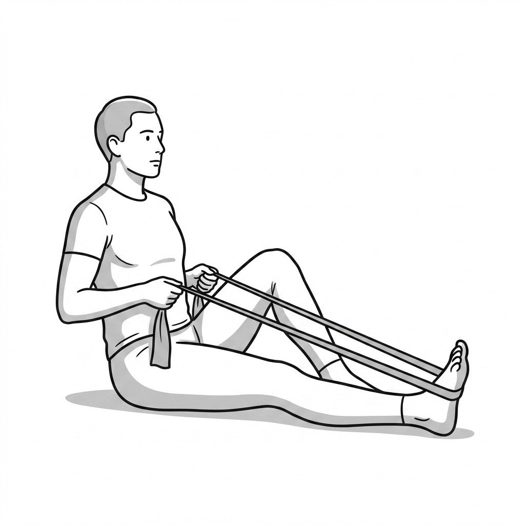
These exercises load the calf-tendon unit through isometric holds and progressive eccentric and heavy-slow work — the combination shown to drive tendon remodelling in both mid-portion and insertional Achilles pathology. Start with 2–3 and progress as tolerated.